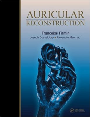
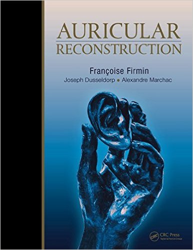
 Authors: Francoise Firmin, MD: Joseph Dusseldorp, MBBS; and Alexandre Marchac, MD
Illustrator: Tiffany Slaybough Davanzo, MA, CMI
Publisher: Thieme – 356 pages
Book Review by: Nano Khilnani
This large-size text with much detailed information and numerous images is a unique one in the medical literature on its subject. There are few if any, other books out there that provide not only step-by-step instructions on ear reconstruction surgery but also a lot of insight developed from decades of experience of the authors.
Dr. Firmin, the lead author of this book, writes in the Preface that he could very well have entitled this book as Three Decades’ Experience in Ear Construction. One of his unique qualifications is that (as he states) he has performed 2,540 procedures on reconstruction of the ear. With this background, he and the co-authors are able to provide you, through the pages of this well-organized, amply-illustrated work:
- Well-established techniques of auricular reconstruction
- Some very fine results of their work
- A realization of how humbling a discipline ear reconstruction is
The authors point out that the ear is a highly complex, three-dimensional anatomic structure with hills and valleys, so to correct partial and total defects, these are some requirements:
- An accurate reproduction of the missing contours and supporting framework
- Principles and guidelines that inform you about the choice of the best support
- Sculpting of the requisite contours
They write that once you adequately understand the three-dimensional structure of the ear, what is then needed by you is mastering soft tissue coverage for the framework. They provide the surgical classification for the skin, which will help determine the appropriate surgical planning for any set of circumstances.
Some of the options available for the support system are:
- Autologous costal cartilage (usually most appropriate for reproducing complex contours)
- Synthetic pre-fabricated framework, mainly to avoid intra-operative carving
The authors cover many subjects within this book. To give you an overview, we provide you the titles of its 17 chapters allocated within six Parts:
- Part I – Two-Stage Autol0gous Auricular Reconstruction
- Principles of Two-Stage Autologous Ear Reconstruction
- Stage I: Reproduction of the Missing Contours
- Stage II: Creation of the RetroAuricular Sulcus
- Part II – Total and Subtotal Defects
- Congenital Malformations
- Particular Cases of Microtia
- Acquired Posttraumatic Defects
- Acquired Defects
- Part III – Management of Unfavorable Conditions
- Secondary Cases
- Complications
- Auricular Prosthesis
- Part IV – Management of Acquired Partial DefectsAnalysis and Principles of Management
- Reconstruction with Conchal and Costal Cartilage
- Particular Cases
- Part V – Minor Anomalies
- Anomalies of the Upper Part of the Ear
- Other Anomalies
- Part VI – Aesthetic Variations
- Prominent Ears
- Complications After Otoplasty
The purchasers of this one-of-a-kind book can also benefit greatly from viewing 18 videos, the titles of which are listed on page xv.
These videos and other valuable items of online content are available at: https://online.vitalsource.com. Two simple steps:
- Create your Vitalsource Bookshelf account
- Redeem the code by scratching off the grey film on the front cover of this book
You can also download the entire E-book edition of this work and read it offline. Detailed instructions are provided at the website address provided above, to download and use the content on your desktop PC or Mac, mobile devices such as iPhone, iPad Touch, or iPad, on your Android, smart phone or tablet, or on your Kindle Fire.
The illustrative assets of this book cannot be overemphasized. Within each chapter, see a large number of different types of ears, among them:
- Bitten ears
- Broken ears
- Concave- and convex-shaped ears,
- Contours that irregular
- Defects on ears
- Discolored ears
- Ears with various anomalies
- Elephant-shaped ears
- Malformed ears
- Oversized ears
- Round-shaped or V-shaped ears
- Undersized ears
Alongside the photos are detailed surgical instructions on how to fix the problems. Sets of photos presented show similarities and differences and provide captions with different options for reconstructive surgical procedures. Before and after-surgery photos are particularly useful.
This is an outstanding and highly valuable and useful resource on auricular reconstruction and its various aspects of surgical treatment and management.
Editors:
Francoise Firmin, MD is a plastic and reconstructive surgeon at Clinic Bizet in Paris, France.
Joseph Dusseldorp, MBBS, MS, FRACS is a clinical lecturer in the Department of Plastic and Reconstructive at the University of Sydney in Sydney, Australia.
Alexandre Marchac, MD is a plastic and reconstructive surgeon at Hospital Europeen Georges-Pompidou and Clinique Bizet in Paris, France.
Illustrator:
Tiffany Slaybough Davanzo, MA, CMI


