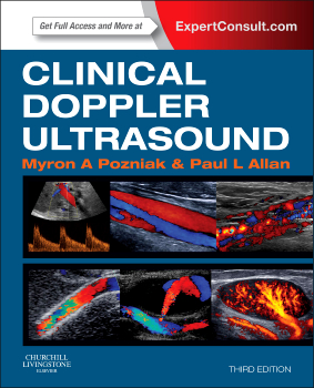
 Authors: Myron A. Pozniak and Paul L. Allan
Publisher: Elsevier Saunders – 390 pages
Book Review by: Nano Khilnani
Doppler ultrasound is a diagnostic method in which measurement and a visual record are made of the shift in frequency of a continuous ultrasonic wave proportional to the blood-flow velocity in underlying vessels; used in diagnosis of extracranial occlusive vascular disease. It is also used in detection of the fetal heartbeat or of the velocity of movement of a structure, such as the beating heart.
The color flow Doppler ultrasound meanwhile is a form of pulse wave Doppler in which the energy of the returning echoes is displayed as an assigned color; by convention echoes representing flow towards the transducer are seen as shades of red, and those representing flow away from the transducer are seen as shades of blue. The color display is usually superimposed on the B-mode image, thus allowing simultaneous visualization of anatomy and flow dynamics.
This book offers you an overview of the applications of Doppler ultrasound in managing patients It helps you distinguish abnormal findings from normal ones, and how to interpret them.
Here are some of the things you can learn to do through this book:
- Understand and apply the latest imaging techniques with a new chapter on interventional and intra-operative applications of Doppler ultrasound and a new chapter on dialysis grafts, plus coverage of the most recent information on the role of contrast agents and how best to administer them.
- View real-time videos of Doppler imaging and search across the complete text online at www.ExpertConsult.com
- Make the most informed imaging decisions possible by gaining a thorough understanding of the advantages and disadvantages of Doppler imaging, as well as the basic principles behind its techniques and technologies.
- Acquire optimal images and avoid errors with the help of detailed protocols and high-quality, full-color illustrations throughout.
The overview below (titles of 17 chapters) of the contents in this book gives you an idea of its coverage and scope. If you’re looking to study on topics not found below, you can go to the lists of References at the end of each chapter.
- Physics: Principles, Practice, and Artefacts
- Haemodynamics and Blood Flow
- The Carotid and Vertebral Arteries; Transcranial Colour Doppler
- The Peripheral Arteries
- The Peripheral Veins
- The Aorta and Inferior Vena Cava
- Haemodialysis Access
- The Liver
- The Kidneys
- Solid Organ Transplantation
- Doppler Imaging of the Prostate
- Doppler Ultrasound of the Penis
- Doppler Imaging of the Scrotum
- Doppler Imaging of the Female Pelvis
- Clinical Applications of Doppler Ultrasound in Obstetrics
- Interventional and Intraoperative Doppler
- Microbubble Ultrasound Contrast Agents
- Appendix: System Controls and Their Uses
Each of the chapters contains study aids to make learning and recall of the material easier. Besides topic headings and discussions, boxes, charts, drawings, figures (of Doppler ultrasound images mainly, but also other types), and tables are provided.
A list of videos that you can view on www.ExpertConsult.com is found in the Video Table of Contents on pages vi and vii of this book
To access the complete contents of this textbook online – which are fully searchable – and to use the other valuable features, simply visit the website indicated above, enter the Activation Code by scratching off the sticker on the inside front cover of this book, and follow the instructions online to activate your access.
If you are already registered, login at www.expertconsult.com, scratch off the Activation Code on the inside front cover of this book, enter it into the Add a Title box, click on Activate Now, and click the title under My Titles.
If you’re a first-time user, click on Register Now, fill in your user information and click Continue, then activate your book by entering your Activation Code into the Enter Activation Code box; then click on Activate Now, and click the title under My Titles.
An additional resource is available to purchasers of this book on www.ClinicalKey.com. Go to the inside back cover of this text, and you will find that ClinicalKey is designed to serve you well by providing you these important core components:
- Comprehensive Content – The most current, evidence-based answers available for every medical and surgical specialty.
- Trusted Answers – Content supplied by Elsevier, the world’s leading provider of health and science information.
- Unrivaled Speed to Answer – Faster, more relevant clinical answers, so you can spend less time searching and more time caring for patients.
This valuable, highly informative, and well-illustrated book with a variety of numerous images was made possible through the expert and tireless efforts of Drs. Pozniak and Allan, with the help of contributors named below.
Myron A. Pozniak, MD, FACR, FSRU, FARS is a Professor of Radiology in the Department of Radiology at the University of Wisconsin School of Medicine and Public Health in Madison, Wisconsin.
Paul L. Allan, BSc, DMRD, FRCR, FRCPE is Consulting Radiologist and Director of Imaging Sciences in the Department of Radiology at Royal Infirmary of Edinburgh in Edinburgh, United Kingdom.
Contributors:
Paul L. Allan, BSc, DMRD, FRCR, FRCPE; Lauren F. Alexander, MD; Jonathan D. Berry. BSC, FRCR; Peter N. Burns, PhD; Michael T. Corwin, MD; Peter R. Hoskins, PGCert, BA, MSc, PhD, DSc; Mark R. Lockhart, MD, MPH; W. Norman McDicken, PhD, FIPEM; John P. McGahan, MD; Imogen Montague, MB ChB, FRCOG, DOU; Fred T. Lee, Jr., MD; Fred Lee, Sr.; Myron A. Pozniak, MD, FACR; Michelle L. Robin, MD; Paul S. Sidhu, BSc, MRCP, FRCR; Heidi R. Umphrey, MD, Therese M. Weber, MD, FACR; and Michael J. Weston, MB ChB, FRCR, MRCP. .