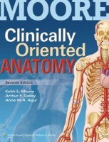
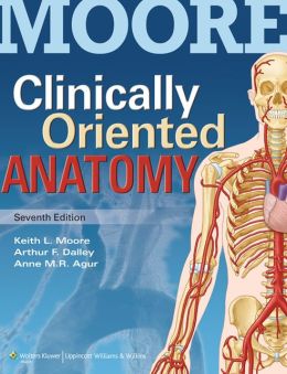
 Authors: Keith L. Moore, Arthur F. Dalley, and Anne M.R. Agur
Publisher: Wolters Kluwer | Lippincott Williams & Wilkins – 1134 pages
Book Review by: Nano Khilnani
This book is different from other texts on anatomy in that it deals with the clinical applications of anatomy. As such, it places clinical emphasis on anatomy. The authors point out that such an approach is important in many activities of physicians, physical or occupational therapists and other health care professionals, including, but not limited to:
- Physical diagnosis for primary care
- Interpretation of diagnostic imaging
- Understanding the anatomical basis of emergency medicine and general surgery.
The first edition came out about a third of a century ago. Anatomy is one of those basic sciences wherein the factual basis does not change, and in this way, this field of study is remarkable for its longevity and consistency.
This edition has many improvements. New imaging technologies have enabled the incredible viewing of living anatomy, and improvements in graphic representation and print publication have made it possible to see details not easily demonstrable before.
This book has seven key features that make it highly useful to students and practitioners:
- Extensive art program – every illustration has been revised, improving accuracy and consistency and giving classical art derived from Grant’s Atlas of Anatomy a fresh, vital, new appearance.
- Clinical correlations – are blue boxes containing clinical information, and many have photographs and / or dynamic color illustrations that help understand the practical value of anatomy.
- Bottom line summaries – enable the reader to understand the important points of information conveyed in the book, and recall them during reviews, in the midst of many details that cannot be remembered.
- Anatomy described in a practical, functional context – the action and use of organs and parts of the body is described. In other words, the functional aspect is given emphasis.
- Surface anatomy and medical imaging – Formerly, surface anatomy and medical imaging were presented separately. Both natural views of unobstructed views of surface anatomy and illustrations superimposing anatomical structures on surface anatomy photographs are components of each regional chapter.
- Case studies, accompanied by clinico-anatomical problems and board review-style multiple-choice questions – for self-testing and review, readers can go to this website: http://thePoint.lww.com to read case studies and questions.
- Terminology – the root words and derivations of terms are provided to help students understand meaning and increase retention. Eponyms appear in parenthesis. The terminology is now available online at
The organization of this book is superb. You see this as soon as you open the book. There are ten tabs on the right, each with a different color, and the edges of the pages have these colors too, so you can easily get to any of the ten sections, which are simply named from top to bottom as: Introduction, Thorax, Abdomen, Pelvis and Perineum, Back, Lower Limb, Upper Limb, Head, Neck, and Cranial Nerves.
This book has many plus points besides the seven key features we enumerated above and its excellent organization of the material. Those plus points are:
- A comprehensive text that enables students to fill in the blanks, as the amount of time they spend in lectures, labs, and studying keeps on increasing.
- A resource that supports areas of special interest within anatomy. This book is good for students of basic anatomical science and those in clinical phases.
- A comprehensive Introduction that provides through summaries of systems within the human body, both descriptive and functional.
- Routine facts are presented in tables organized to demonstrate shared qualities and illustrated to demonstrate the provided information.
- Illustrated clinical correlations that show anatomy applied clinically; graphics that facilitate orientation, such as arrows, labels, locations and viewing sequences
- Blue boxes classified by following icons indicating type of clinical information covered: Anatomical variations, Life cycle, Trauma, Diagnostic procedures, Surgical procedures, and Pathology.
There are hundreds of multi-dimensional, full-color drawings throughout out the book to enable the reader to take detailed looks at systems and organs from many perspectives. Some X-rays are also provided that show abnormalities that can be compared with the full-color drawings that show normal views of the particular part of the body in question.
This is an excellent book with information that is ample and comprehensive and very well presented for easier learning and retention. The authors have done a marvelous job and their contributions to the advancement and effective dissemination of anatomical knowledge are no less short of outstanding.
Keith L. Moore, Ph.D., M.Sc., D.Sc. is professor emeritus in the division of anatomy in the department of surgery, former chair of anatomy, and associate dean for basic medical sciences at the faculty of medicine at the University of Toronto in Ontario, Canada.
He is the recipient of many prestigious awards and recognitions. He has received the highest awards for excellence in human anatomy education at the medical, dental, graduate and undergraduate levels – and for his remarkable record of textbook publications in clinically oriented anatomy and embryology – from both the American Association of Anatomists (AAA: Distinguished Education Award, 2007) and the American Association of Clinical Anatomists (AACA: Honored Member Award, 1994)
Arthur F. Dalley II, Ph.D. is professor in the department of cell and development biology, adjunct professor in the department of orthopedics and rehabilitation, and director of programs in medical gross anatomy and the anatomical donations program at the Vanderbilt University School of Medicine. He is also adjunct professor of anatomy at the Belmont University School of Physical Therapy in Nashville, Tennessee.
Anne M.R. Agur, B.Sc. (OT), M.Sc., Ph.D. is on the faculty of medicine at the University of Toronto in Ontario, Canada. She is professor in the division of anatomy in the departments of surgery, physical therapy, occupational science and occupational therapy. She also works in the division of physiatry at the university’s department of medicine; at the division of biomedical communications at the institute of medical science; at the graduate department of rehabilitation science and at the graduate department of dentistry.


