 Editors: Amar Agarwal, FRCOphth and Dhivya Ashok Kumar, MD
Publisher: Thieme – 245 pages
Book Review by: Nano Khilnani
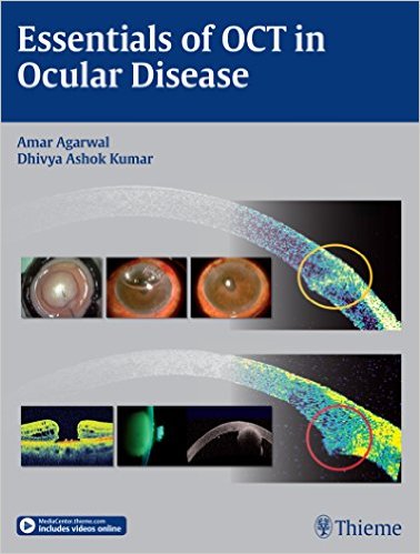
Ocular coherence tomography (OCT) is a relatively newer method of investigating eye disorders. It is now widely used by ophthalmologists and optometrists to perform high-resolution, micrometer-scale, cross-sectional imaging of ocular tissues in less time than before. OCT machines, such as the ones shown on page 4, 17 and 53 of this book, enable practitioners to do diagnosis, prognosis and management of ocular pathologies.
OCT as a form of imaging utilizes low-coherence interferometry to create a two-dimensional image of optical scattering from internal tissue microstructures. In a way it is similar to ultrasonic pulse-echo imaging.
This imaging modality is better than others such as computed tomography (CT), magnetic resonance imaging (MRI), and ultrasound, because of various reasons, among them:
- Its noninvasive nature
- Its higher resolution
- Its versatility of use
- Its wide acceptance as one of several standard diagnostic tests for many ocular disorders
Indeed, OCT has become the imaging modality of choice for eye care professionals. It was first used for in vitro imaging in the peripapillary area of the retina and in the coronary artery. The former being a transparent area and the latter a turbid one, OCT has proven its adaptability.
Fifty-two practitioners (physicians and surgeons), professors, and researchers in optometry, ophthalmology and related fields authored the 28 chapters that constitute this volume. The material is organized around 5 Parts, listed below. The writers are from: Argentina, England, France, Germany, Hong Kong, India, Saudi Arabia, South Korea, the United States, and Venezuela.
- Basics: chapters 1 to 5
- Anterior-Segment Optical Coherence Tomography: chapters 6 to 13
- Cataract Surgery: chapters 14 to 18
- Optical Coherence Tomography in Retinal Diseases: chapters 19 to 22
- Miscellaneous: chapters 23 to 28
The purchasers of this book can view 10 highly useful videos we have listed below. Simply visit www.MediaCenter.Thieme.com and when prompted during the registration process, enter the access code found on the inside front cover of this book.
Video 1: Room with a View
Video 2: Corneal Biomechanics: The New Mantra
Video 3: Posterior Chamber Phakic IOL: The Right Way Up
Video 4: Contact Lens Assisted Collagen Crosslinking in Thin Corneas: New Technique
Video 5: Descemet’s Detachment New Classification and Management
Video 6: Pre-Descemet’s Endothelial Keratoplasty
Video 7: Sub 1 mm Cataract Surgery
Video 8: Glue IOL Reloaded
Video 9: What Lies Beneath
Video 10: Stab Incision Glaucoma Surgery
Simple yet skillful organization of material in the chapters by authors and editors enable readers to learn and comprehend the subjects more easily, and better retain the information for practice and later reference. Chapters typically have these parts:
- Background or History
- Main Topics
- Subtopics
- Conclusion
- References
Numerous visuals using various imaging modes are found in this book, among them:
- Angiography
- Charts
- Fundus photography
- OCT scanning
- Tables
This is an excellent source of information on how to use ocular coherence tomography or OCT to examine the structures and functions of the eye, detect diseases and disorders, plan and implement treatment.
Editors:
Amar Agarwal, MS, FRCS, FRCOphth is Professor and Chairman and Managing Director of Dr. Agarwal’s Group of Eye Hospitals and Eye Research Centre in Chennai, Tamil Nadu, India. He is Past President of the International Society of Refractive Surgery and Secretary General of the Indian Intraocular Implant and Refractive Society.
He is the pioneer of phakonit (phako with needle incision) technology. This technique became popular as bimanual phaco, microincision cataract surgery, or microphaco. He was the first to remove cataracts through a 0.7-mm tip using a technique called microphakonit and initiated no-anesthesia cataract surgery and sleeveless phacotip-assisted levitation, previously known as FAVIT (fallen vitreous), a new technique to remove dropped nuclei.
Dhivya Ashok Kumar, MD, FICO is Consultant Ophthalmologist and Head of Research and Development at Dr. Agarwal’s Group of Eye Hospitals and Eye Research Centre in Chennai, Tamil Nadu, India. Dr. Kumar completed her ophthalmology residency training at the All India Institute of Medical Sciences in New Delhi and has been affiliated with Dr. Agarwal’s Eye Hospital and Eye Research Centre since 2007.
She is now working as a consultant in uvea and oculoplasty services at Dr. Agarwal’s Eye Hospital. She has an immense passion for research and innovative methods for the evaluation of ocular disorders. Her regions of interest include anterior-segment imaging, glued intraocular lens, and ocular inflammation.