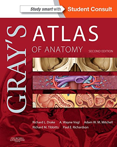
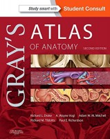
 Authors: Richard L. Drake, PhD; A Wayne Vogl, PhD; and Adam W.M. Mitchell, MBBS
Illustrators: Richard M. Tibbits and Paul E. Richardson
Publisher: Elsevier Saunders – 626 pages
Book Review by: Nano Khilnani
This is a companion resource to the popular Gray’s Anatomy for Students. It in you will find accurate and clear depictions of the anatomy of eight regions of our bodies, as presented in the outline of contents of this book:
- The Body
- Back
- Thorax
- Abdomen
- Pelvis and Perineum
- Lower Limb
- Upper Limb
- Head and Neck
To get the most from this book, get the online resources available to you to Study Smart with Student Consult at https://studentconsult.inkling.com/ Here’s what you do:
- Login and sign up at the above site
- Scratch off your PIN code found on the inside front cover of this book
- Enter your PIN on the Redeem a Book Code box
- Click on Redeem
- Go to My Library
With your personal computer (PC), Mac, most mobile devices, and eReaders, you can browse, search, and interact with this title, online and offline. Here are some of the neat features of this online resource:
Seamless, real-time integration between devices
- Straightforward navigation and search
- Notes and highlights: interactive sharing with other users through social media
- Adjustable text size and brightness
- Enhanced images with annotations, labels, and hot spots for zooming on specific details*
- Live streaming video and animations*
- Self-assessment tools: questions embedded within the text and multiple-format quizzes*
*- Some features vary by title
This extensive and intensive atlas of over 600 pages provides detailed full-color drawings of anatomical structures, and images obtained through computed tomography (CT) and magnetic resonance (MR) technologies. Many of the CT images were taken with contrast, so they present the anatomy in question much more clearly than would be possible in simple CT depictions.
This updated second edition contains a lot of new material. Among other benefits, you will derive the following from this version:
- Demonstrates the correlation of structures with appropriate clinical images and surface anatomy essential for proper identification in the dissection lab and successful preparation for course exams.
- Makes it easier than ever to master the essential anatomy knowledge you need, with its clinical focus, consistent and clear illustrations, and logical organization of material.
- Provides summary tables and schematic diagrams at the end of each chapter that cover relevant arteries, muscles, and nerves,
- Gives you full access to the entire book contents, plus dissection video clips, and self-assessment questions at www.studentconsult.com.
Gray’s Atlas of Anatomy, second edition is an excellent resource for students and teachers of human anatomy. The fact that so much additional information is available and interaction is possible online makes it a truly outstanding, highly valuable product.
Authors:
Richard L. Drake, PhD, FAAA is Director of Anatomy and Professor of Surgery in the Cleveland Clinic Lerner College of Medicine at Case Western Reserve University in Cleveland, Ohio.
A Wayne Vogl, PhD, FAAA is Professor of Anatomy and Cell Biology in the Department of Cellular and Physiological Sciences Faculty of Medicine at the University of British Columbia in Vancouver, British Columbia, Canada.
Adam W.M. Mitchell, MBBS, FRCS, FTCT is Consultant Radiologist and Senior Lecturer at Imperial College at Chelsea and Westminster Hospital in London, UK.
Foreword by: Susan Stranding, PhD, Disc, FKC, Hon FRCS – Emeritus Professor of Anatomy at King’s College in London.
Illustrators:
Richard M. Tibbits
Saffron Walden, UK
Paul E. Richardson
Cambridge, UK
Photographer:
Ansell Horn

