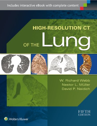
 Authors: W. Richard Webb, MD, Nestor L Muller, MD, and David P. Naidich, MD
Contributing Authors: Brett M. Elicker, MD, Myrna C.B. Godoy, MD, and C. Isabella S. Muller, MD.
Publisher: Wolters Kluwer | Lippincott, Williams & Wilkins – 732 pages
Book Review by: Nano Khilnani
High-Resolution Computed Tomography (HRCT) has become an established tool for evaluating patients with diffuse lung disease, over the last 25 years since the first edition of this book appeared in 1991.
Much progress has also occurred in the understanding of diffuse lung diseases in the last five years since the fourth edition of this book appeared. Documenting all that progress and the new discoveries warranted the publication of this current edition.
This latest – fifth – edition covers a broad spectrum of lung abnormalities, ailments, diseases, and disorders, as you can see from the titles of its 24 chapters in four sections below:
- High-Resolution CT Techniques and Normal Anatomy
- Technical Aspects of High-Resolution CT
- Normal Lung Anatomy
- Approach to HRCT Diagnosis and Findings of Lung Disease
- HRCT Findings: Linear and Reticular Opacities
- HRCT Findings: Multiple Nodules and Nodular Opacities
- HRCT Findings: Parenchymal Opacification
- HRCT Findings: Air-Fluid Cystic Lesions
- HRCT Findings: Decreased Lung Attenuation
- High-Resolution CT Diagnosis of Diffuse Lung Disease
- Idiopathic Interstitial Pneumonias, Part I: Usual Interstitial Pneumonia/Idiopathic Pulmonary Fibrosis and Nonspecific Interstitial Pneumonia
- Idiopathic Interstitial Pneumonia, Part II: Cryptogenic Organizing Pneumonia, Acute Interstitial Pneumonia, Respiratory Bronchiolitis-Interstitial Lung Disease, Desquamative Interstitial Pneumonia, Lymphoid Interstitial Pneumonia, and Pleuroparenchymal Fibroelastosis
- Collagen-Vascular Diseases
- Diffuse Pulmonary Neoplasms and Pulmonary Lymphoproliferative Diseases
- Sarcoidosis
- Pneumoconiosis, Environmental and Occupational Lung Disease
- Hypersensitive Pneumonitis and Eosinophilic Lung Diseases
- Drug-Induced Lung Diseases and Radiation Pneumonitis
- Miscellaneous Infiltrative Lung Diseases
- Infections
- Pulmonary Edema and Acute Respiratory Distress Syndrome
- Cystic Lung Diseases
- Emphysema and Chronic Obstructive Pulmonary Disease (COPD)
- Airways Diseases
- Pulmonary Hypertension and Pulmonary Vascular Disease
- High-Resolution CT Review
- Illustrated Glossary of High-Resolution CT Terms
- Appearances and Characteristics of Common Diseases
While going through the print edition will provide you a lot of detailed knowledge, you can benefit even more when you go online. With your purchase of the print edition of this text, you can enjoy its bundled interactive eBook edition as well, offering tablet, smartphone and online access to:
- Complete content with enhanced navigation that can be accessed anywhere, even without a data connection
- Powerful search and smart navigation cross-links
- Highlighting tool for easier reference of key content throughout the text
- Ability to take and share notes with friends and colleagues
- Quick reference tabbing to save your favorite content for future use
Here’s how you download your eBook:
- Go to http://solution.lww.com/access
- Enter the Access Code found by scratching off the tab on inside front cover of your book
- Sign in or create an account to complete checkout and start reading.
The coverage in this book of diffuse lung diseases is not only extensive but also intensive to some extent, going into detail with the disease or diseases each chapter is focused on.
For example chapter 12 entitled Sarcoidosis discusses the pathologic and radiologic findings of this disease, but also the associated conditions and sarcoid-like reactions. The chapter also provides a differential diagnosis of sarcoidosis, and numerous high-resolution CTs are presented in the chapter.
Especially instructive among the images shown is Figure 12-21 C which depicts bronchial involvement in a patient having sarcoidosis. The caption states: “multiple endobronchial nodules (arrows) represent granulomas.” Actually seeing a disease beats reading about it, even if explained in detail in words. Needless to say, a picture is worth a thousand words. And this book is chock full of images that makes learning easier and quicker.
Tables provide data at a glance from differential diagnoses to understand the details of diseases, and lists of reference materials point out to other sources of information when you are trying to find answers to questions in a given case. This is a valuable book written by six physicians who are expert in imaging and treating lung diseases, who we name below.
Authors:
W. Richard Webb, MD is Professor Emeritus of Radiology and Biomedical Imaging; and Emeritus Member of the Haile Debas Academy of Medical Educators at the University of California San Francisco in San Francisco, California.
Nestor L Muller, MD, PhD is Professor Emeritus of Radiology in the Department of Radiology at the University of British Columbia in Vancouver, British Columbia, Canada.
David P. Naidich, MD, FACR, FAACP is Professor of Radiology and Medicine at New York University Langone Medical Center in New York, New York.
Contributing Authors:
Brett M. Elicker, MD is Associate Professor of Clinical Radiology and Biochemical Imaging, and Chief of Cardiac and Pulmonary Imaging at University of California San Francisco in San Francisco, California.
Myrna C.B. Godoy, MD, PhD is Associate Professor of Radiology at University of Texas MD Anderson Cancer Care Center in Houston, Texas.
C. Isabella S. Muller, MD, PhD is with the Department of radiology at Delfin Clinic in Salvador, Bahia, Brazil.