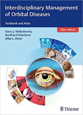
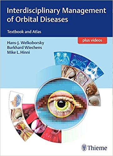
 Editors: Hans-J. Welkoborsky, MD; Burkhard Wiechens, MD; and Michael L. Hinni, MD:
Publisher: Thieme – 340 pages, with 689 Illustrations, plus videos
Book Review by: Nano Khilnani
The orbit is the bony orifice region of the human head where the eye and its appendages are situated. The appendages include the eye socket, eyelids, eyebrows, the lacrimal apparatus, conjunctiva, and the associated blood vessels, bones, glands, and muscles.
The orbit is a small, bony compartment with thin bony borders towards the paranasal sinuses, and it is a busy thoroughfare of pathways to and from the face. In describing this region, A. Schmeid, author of chapter 1 – Topographical Anatomy of the Orbit – writes: “significant hemorrhage can occur during operative procedures” due to the presence of numerous blood vessels in this field” This area has 10 branches of the ophthalmic artery, two large veins, and their afferent vessels.
Twenty-nine specialists in craniomaxillofacial surgery, otorhinolaryngology – head and neck surgery, and related fields such as ophthalmology, and a variety of other medical areas such as anesthesiology, cell and molecular physiology, functional and applied anatomy, neurology, neuro-radiology, neurosurgery, and radiation oncology, authored the 19 chapters of this large-size (12’’x 9”) book.
The contributors are mainly based in Germany, but they are also in three other countries – Spain, Switzerland, and the United States. We list the chapters of this book below to give you a broad overview of its coverage:
- Topographical Anatomy of the Orbit
- Pathophysiological Aspects of the Orbit
- Clinical Examination in Patients with Orbital Diseases
- Imaging of the Orbit
- Diseases of the Eyelids and the Eye
- Orbital Complications
- Endocrine Orbitopathy
- Traumatology and Traumatic Optic Neuropathy
- Pathology of the Orbit
- Orbital Malformations
- Tumors of the Orbit and the Eye
- Tumors of the Fronto-Orbital Skull Base, the Optic Nerves, and the Intracranial Optic Pathways
- Intraconal Orbital Tumors
- Radiotherapy of Orbital Diseases
- Surgery of the Lacrimal Drainage System
- Lid Surgery in Orbital Disorders
- Anesthesiological Aspects of Orbit Surgery
- Surgical Approaches to the Orbit
- Reconstructive Orbital Surgery
Videos demonstrating the surgical procedures covered in this book are available on www.MediaCenter.Thieme.com, where you simply enter the code found on the inside front cover of this book, to gain access.
A closer look at the anatomical structures within the orbit as found in chapter 1, takes you to detailed discussions and fine-line illustrations of the following:
- Introduction
- Shape, Borders, Orifices, and Topographical Relations
- Bony orbital Walls
- Orbital Foramina
- Topographical Relations of the Orbit
- Protective Apparatus of the Eye
- Oribital Septum
- Eyelids
- Microanatomy and Blood Supply
- Locomotor System of the Eyelids: Lid Muscles
- Striped Muscles
- Unstriped Muscles
- Palpebral Limbus and Palpebral Glands
- Lacrimal Apparatus
- Contents of the Orbit
- Classification
- Connective Tissue
- Periorbita
- Corpus Adiposum
- Vagina bulbi
- Fibrous Suspension Apparatus
- Pars Bulbosa and Bulbus Oculi
- Layers of the Eyeball (Eyeball Tunica)
- Eyeball Compartments
- Eye Lens and Suspension Apparatus
- Topography and Function of Outer Eye Muscles
- Layers of the Eyeball (Eyeball Tunica)
- Topography of Pathways Located Postseptally
- Topographic Relations to the Common Anular Tendon of Zinn
- Extraconal Pathways in the Upper Compartment
- Nerves and Arteries
- Veins
- Intraconal Pathways (Medial Compartment)
- Nerves
- Ophthalmic Artery and Its Branches
- Veins
- 1.5.4 Extraconal Pathways (Lower Compartment)
- Nerves and Arteries
- Veins
- 1.5.5 – Secretory, Parasympathetic Innervation of the Lacrimal Gland
References
This unusually well-illustrated book – Interdisciplinary Management of Orbital Diseases – Textbook and Atlas – has the following noteworthy characteristics:
Key Features:
- Valuable guidance from leading specialists in craniomaxillofacial surgery, endocrinology, neurosurgery, ophthalmology, otorhinolaryngology, and other relevant fields
- Coverage of the spectrum of orbital diseases and pathologies, including congenital malformations and posttraumatic deformities orbital and skull base tumors and inflammation, sinus complications, thyroid eye disease, and more
- Useful details on patient examination, surgical techniques and pitfalls, and postoperative management aid in daily practice
- More than 650 full-color photographs and illustrations that allow for rapid learning and application of concepts
- Videos accessed through Thieme MediaCenter that enhance understanding of surgical procedures
This is an outstanding volume with excellent, extensive coverage, through words and hundreds of images, of the anatomy and physiology of the orbital region of the head, including diagnosis and medical and surgical treatment of diseases in this region.
Editors:
Hans-J. Welkoborsky, MD, DDS, PhD is Professor and Chairman in the Department of Otorhinolarygology – Head and Neck Surgery at Norstadt Clinic, Academic Hospital and Children’s Hospital “Auf dur Bult” in Hannover, Germany.
Burkhard Wiechens, MD, PhD is Professor and Chairman of the Department of Ophthalmology at Norstadt Clinic, Academic Hospital in Hannover, Germany.
Michael L. Hinni, MD is Professor and Chairman in the Department of Otolaryngology – Head and Neck Surgery at Mayo Clinic in Scottsdale, Arizona.


