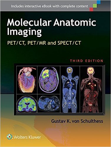
 Editor: Gustav K. von Schulthess, MD
Publisher: Wolters Kluwer – 683 pages
Book Review by: Nano Khilnani
Molecular imaging is not a new development. It has been around about 70 years. In 1946 the Journal of the American Medical Association reported on targeted imaging of iodine therapy for metastatic thyroid cancer.
This third edition provides new information and findings, and is an update to the second edition that appeared in 2007. Since that time, hybrid imaging techniques have become quite commonplace, with numerous important technological developments
Hybrid imaging on a molecular level is imaging using a combination of two or more imaging techniques, such as positron emission tomography (PET) and computed tomography (CT). Another type of hybrid imaging is single-photon emission computed tomography (SPECT) with CT. Hybrid imaging is now used about eighty percent of the time when imaging is required, writes Dr. Schulthess in the Preface.
He adds that this hybrid use is true all over the world, even in less developed countries. And as this book has been published in 2015, hybrid PET with magnetic resonance (MR) imaging systems are now being introduced.
A common component of all types of hybrid imaging is the use of radioactive materials or tracers, including pharmaceuticals.
One hundred and five MDS and PhDs from all over Europe – Austria, Denmark, England, Germany, Italy, the Netherlands, Sweden, Switzerland – and the United States and Canada, as well as Hong Kong, Israel, Singapore, and South Korea, wrote the 73 chapters of this book. It is organized into seven Parts as shown below:
- Basic Aspects of Hybrid Imaging Technology (chapters 1 to 12)
- Radiopharmaceuticals (chapters 13 to 16)
- Clinical Hybrid Imaging of the Brain (chapters 17 to 26)
- Clinical Multimodality Imaging of the Heart ( chapters 27 to 30)
- Clinical Hybrid Imaging in Body Oncology:
- Nonneoplastic Disease in the Body (chapters 63 to 70)
- Clinical Hybrid Imaging in Pediatric Patients (chapters 71 to 73)
The entire contents of the print edition are available for download as an eBook. Follow these simple steps:
- Go to http://solution.lww.com/access
- Enter the Access Code found on the inside front cover of this book
- Enter your information, click Submit, and follow the on-screen instructions to start reading your eBook
Your book purchase includes not only a complimentary download of the enhanced eBook for iOS, Android, PC and Mac, but also these features:
- Complete content with enhanced navigation
- Powerful search tools and smart navigation cross-links that pull results from content in the book, your notes, and even the web
- Cross-linked pages, references, and more for easy navigation
- Highlighting tool for easier reference of key content throughout the text
- Ability to take and share notes with friends and colleagues
- Quick reference tabbing to save your favorite content for future use
In the Foreword, Hedvig Hrecak points out that we live in one of the most exciting times today, with not only new discoveries happening rapidly in medicine, but also the convergence of life science, physical science, and engineering. He writes that in the last 30 years, new developments in computer science, engineering and physics have led to new biomedical imaging capabilities.
But what is even more useful is the concerted effort to not only integrate the various imaging technologies to provide better diagnosis, but also to incorporate chemistry and biology to provide more precise, localized, and better-illuminated detection of disease. Better detection provides the basis for correct diagnosis which in turn leads to more effective treatment and well-being.
Since 2007, radiopharmaceutical developments and their clinical applications have expanded significantly. In the past, clinical PET was dominated by Fludeoxylglucose (FDG). Some of the new tracers being used nowadays are: amino acids, (brain) amyloid imaging agents, and choline. The current edition provides a lot of information on current tracers and ongoing developments in this area.
This book is an outstanding and comprehensive contribution to expanding the knowledge base on precision imaging, which is so crucial in the diagnosis and treatment of disease.
Editor:
Gustav K. von Schulthess, MD, PhD, MD hon is Professor, Chairman and Director Emeritus of the Department of Medical Radiology and Nuclear Medicine at University Hospital Zurich in Zurich, Switzerland.