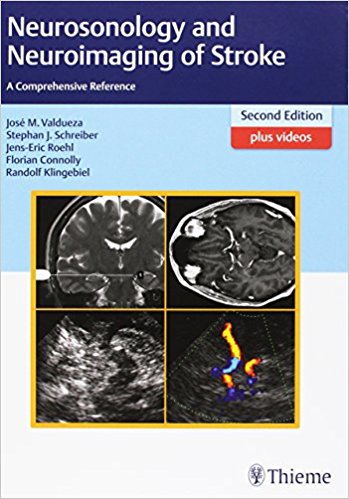
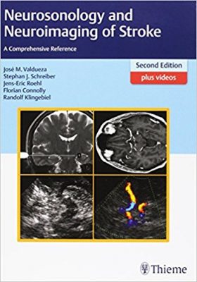
 Authors: Jose M. Valdueza, MD; Stephen J. Schreiber, MD; Jens-Eric Roehl, MD; Florian Connolly, MD; and Randolf Klingebiel, MD
Publisher: Thieme – 619 pages
Book Review by: Nano Khilnani
Stroke occurs when a blood vessel in the brain is blocked, but the exact and detailed causes of the blockage cannot be easily determined until a thorough examination is conducted, the authors of this book point out in the Preface.
The machines used since the 1970s to look at the extent and other characteristics of blood constriction have been of several types, with varying capabilities. One of the first technologies used to view brain-supply vessels was conventional angiography.
Later, in the 1980s, computed tomography (CT) and CT angiography scanning were developed, and were (as they still are) widely used. So is the case of magnetic resonance imaging (MRI).
But CTs and MRIs have limitations. CT involves exposure to radiation and CTA requires the insertion into the veins of a contrast agent; and MRI cannot be used for patients on a pacemaker.
Today one the best imaging modalities is diagnostic ultrasound because it can be used quickly and spontaneously. Among its advantages: it is dynamic, inexpensive, noninvasive, and powerful, able to present clear three-dimensional views of the vessels.
Ultrasound follows rules and pathways, but it also enables the physician, along with the sonographer perhaps, to be at the patient’s bedside, communicate with him or her, and make real-time decisions right then and there on the next course of action.
This thick book on examining stroke though a variety of imaging modalities takes a practical approach by looking at actual cases, after the basic principles are presented and discussed, as we show you in the outline of its contents below:
- Part A. Principles and Rules
- Flow and Ultrasound Basics
- Vascular Anatomy and Structure of Ultrasound Examination
- Intracranial Hemodynamics and Functional Tests
- Pathogenesis of Stroke
- Vascular Pathology
- Angiographic Techniques in Neuroradiology
- Part B. Case Histories
- Cases are presented in this section, ranging alphabetically from ‘ascending left middle cerebral artery occlusion in an HIV positive patient’ to ‘subclavian steal in left subclavian artery and right internal carotid artery occlusion leading to extracranial–intracranial bypass surgery.
In addition to contents in your printed book, you can also access videos and ultrasound reference charts online at: www.MediaCenter.Theme.com. To get your access code, simply scratch off the gray film found on the inside front cover of your book. When prompted while registering your book, type in your access code.
Here are key features of this text:
- Complete extra-cranial and intracranial arterial and venous ultrasound examination
- New techniques: ultrasound fusion imaging, ultrafast ultrasound, contrast application
- Fifteen newly selected cases on conditions such as subarachnoid hemorrhage and dural fistula, as well as rare stroke causes including sickle cell disease and reversible cerebral vasoconstriction syndrome
- More than 60 new video clips (for a total of 130) available in the Thieme Media Center, bringing ultrasound anatomy and challenging cases to your monitor!
Vascular Anatomy and Structure of Ultrasound Examination is the title of chapter 2, which is a core part of this highly useful book on imaging. Consisting of about 60 pages packed with lots of detailed information and illustrations, this chapter covers numerous aspects of arterial and venous anatomy, and how ultrasound diagnostics helps us to view and understand them. It includes following topics:
- General Arterial Anatomy
- Extra-cranial and Intracranial Arterial Anatomy
- General Structure of Arterial Ultrasound Examination
- Special Arterial Anatomy and Ultrasound Anatomy
- Extra-cranial and intracranial Arteries
- General Venous Anatomy
- Extra-cranial and Intracranial Venous Anatomy
- General Structure of Venous Ultrasound Examination
- Special Venous Anatomy and Ultrasound Anatomy
- Extra-cranial and Intracranial Veins
This second edition of Neurosonology and Neuroimaging of Stroke offers an excellent overview of the basic principles of neurosonology. In it the five authors of this book present and discuss the advantages and limitations, as well as the clinical and technical applications of ultrasound as they relate to cerebrovascular anatomy and physiology, particularly stroke.
They compare and contrast ultrasound, a relatively newer imaging technology, with computed tomography or CT, as well as CT angiography (CTA) and magnetic resonance imaging, or MRI. A review of 45 patient histories including diagnoses of their conditions makes the book down-to-earth and connected to reality. This is probably the best new and updated book available on the neurosonology and neuroimaging of stroke to add to your medical library.
Authors :
Jose M. Valdueza, MD is Professor of Neurology at Charite – Universitatsmedizin Berlin in Berlin, Germany, and Director of the Center of Neurology at Segeberger Kniken Gruppe in Bad Segeberg, Germany.
Stephen J. Schreiber, MD is Professor of Neurology at Charite – Universitatsmedizin Berlin in Berlin, Germany, and Head of the Department of Neurology at Asklepios Hospital Brandenbergan der Havel, Germany.
Jens-Eric Roehl, MD is Senior Neurologist in the Department of Neurology at Klinikum Ernst von Bergmann in Potsdam, Germany.
Florian Connolly, MD is Senior Neurologist in the Department of Neurology at Universitatsmedizin Berlin in Berlin, Germany.
Randolf Klingebiel, MD is Professor of Neuroradiology at Universitatsmedizin Berlin in Berlin, Germany, and Head of the Department of Diagnostic and Interventional Neuroradiology at Evangelisches Krankenhaus Bielefeld in Bielefeld, Germany.