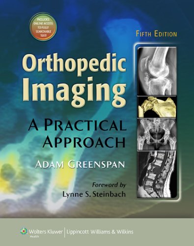
 Author: Adam Greenspan, MD, FACR
Publisher: Wolters Kluwer | Lippincott, Williams & Wilkins – 986 pages
Book Review by: Nano Khilnani
This massive work of nearly a thousand pages is a wonder at first sight! Upon opening it, I could not find a list of contributors, as is the case with books of this size and even smaller ones. Not only does it contain a huge amount of information including more than 4,000 high-quality images, it is mainly the work of one person: the internationally-renowned and eminent musculoskeletal radiologist Dr, Adam Greenspan. He is not just the editor; he is the sole author of this large but excellent text in many respects.
Upon reading up a variety of views and reviews on this book, I discovered that this is a long-standing, popular, extensive, authoritative, and respected text on the imaging of bones and muscles. It’s been written for reference and use by radiologists and orthopedists, no matter what level of knowledge and training they are at.
Orthopedic Imaging: A Practical Approach covers a large range of orthopedic problems. It also shows examples of many types of musculoskeletal imaging, including those that are useful in:
- arthritis
- congenital and developmental anomalies (including a variety of dysplasia, endocrine, metabolic and systemic diseases, infections and neoplasms of the musculoskeletal system including spine)
- sports medicine
- trauma
One of the many valuable benefits it provides is guidance on what and how to select imaging techniques based on cost effectiveness.
This latest – fifth – edition has been expanded and revised. It contains many changes and improvements over the previous one published six years ago. Per Dr. Lynne Steinbach who wrote the Foreword, this fifth edition:
- includes the use of the newest imaging modalities, while remaining as a single volume
- has a wide-ranging look at musculoskeletal imaging that includes magnetic resonance imaging (MRI), computer tomography (CT), three-dimensional (3D) imaging, nuclear medicine imaging (scintigraphy), positron emission tomography (PET scan), ultrasound and radiographs.
- also covers procedures that are more commonly performed by musculoskeletal radiologists – arthrography, percutaneous image-guided biopsy, and radio frequency ablation – are also covered.
- stresses image guidelines throughout the text
- mentions whenever appropriate, therapeutic approaches, pathology, and cytogenetics for musculoskeletal disease
Additionally, this fifth edition has a new full-color design, with colorized tables and schematics and full-color illustrations.
This text comes with access to the complete contents online, full searchable, plus other valuable features. Scratch off the gray sticker on the inside front cover of your book to get your access code. Three simple steps to gain instant access:
1) Visit http://solution.lww.com
2) Enter your access code
3) Follow the instructions to activate your access
For technical assistance, call 1-800-468-1128 in the United States or 1-410-528-4000 outside the U.S., or email: [email protected]
This book contains an extensive coverage of subjects through its 33 chapters, organized within seven Parts entitled:
- Introduction to Orthopedic Imaging
- Trauma
- Arthritides
- Tumor and Tumor-Like Lesions
- Infections
- Metabolic and Endocrine Disorders
- Congenital and Developmental Anomalies.
Each chapter contains a large number and variety of musculoskeletal images, plus other study aids such as a range of different graphics in the Figures and Tables, to make the absorption of material easier for the reader. A very helpful feature at the end of each chapter, for students to read, respond, record and review (part of the SQ4R method in which the first letters stand for Survey and Question) is the Practical Points to Remember section.
This book has numerous useful features that make it highly valuable. This is truly a monumental work of the author. Therefore it is really no wonder that it is a widely-used text on musculoskeletal imaging
Author: Adam Greenspan, MD, FACR is Professor Emeritus of Radiology and Orthopedic Surgery at the University of California, Davis School of Medicine. He is Former Director of the Section of Musculoskeletal Imaging in the Department of Radiology of the UC Davis Medical Center in Sacramento, California. He is also a Consultant to the Shriners Hospital for Children in Sacramento, California.
Foreword by: Lynne S. Steinbach, MD, FACR is Professor of Radiology and Orthopedic Surgery and Director of Musculoskeletal Imaging in the Department of Radiology at the University of California in San Francisco, California.


