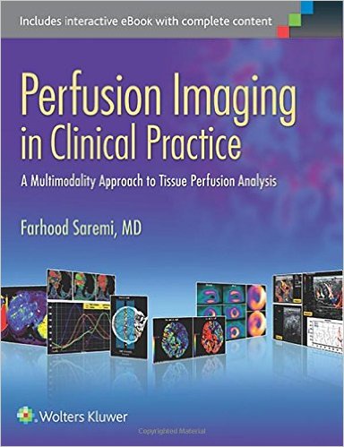
 Editor: Farhood Saremi, MD
Publisher: Wolters Kluwer | Lippincott, Williams & Wilkins – 592 pages
Book Review by: Nano Khilnani
Perfusion in medical science is the flow of blood or other fluid through or over tissue in an organ. Imaging is the viewing of such flow through a scanning machine, device or instrument.
A common example of perfusion is the myocardial nuclear perfusion test used to determine if there is insufficient blood in the heart and / or if there is a blockage in blood vessels. A small amount of thallium-201 or other radioactive substance (sometimes termed ‘contrast’) is injected into the veins of the patient, and images are then produced by a scanner, first at rest and later, after running on a treadmill.
The presence or absence of blockages in the form of fatty deposits (sometimes called ‘plaque’) on the walls of the blood vessels help determine if the patient has or does not have coronary artery disease, the leading cause of death in the United States today. The narrowing of the vessels reduces blood flow to the heart muscle. An insufficient blood supply to the heart is caused by the narrowing of the blood vessels.
This is often manifested in symptoms such as chest pain and shortness of breath, resulting from reduced oxygen that is carried through the blood.
The purpose of this book is to show and measure tissue perfusion in different organs, and to describe the various approaches to imaging that perfusion, states the editor Dr. Farhood Saremi.
The clinical applications of these techniques of perfusion and imaging are vast and quite broadly scattered in medical literature, the editor points out. This makes it quite difficult for radiologists and clinicians to understand the practical clinical implications of these diagnostic methods.
Eight-eight doctors of medicine and fields related to perfusion and imaging, from the United States and 13 other countries – Australia, Canada, Denmark, France, Germany, Greece, Italy, Japan, the Netherlands, Saudi Arabia, Spain, Switzerland, the United Kingdom – contributed material for this book, mainly writing its 35 chapters, which we present below to give you an overview of what you will (or will not) find in this volume:
- Vascular Anatomy and Microanatomy
- Computed Tomography (CT) Perfusion Imaging
- New Dose Reduction Strategies in Myocardial Perfusion Imaging
- Dynamic Contrast-Enhanced Perfusion CT
- CT and MR Contrast-Enhanced Tissue Perfusion Imaging
- Coronary Fractional Flow Reserve Based on Computed Tomography
- Dynamic Susceptibility Contrast MRI: Technical Considerations with Practical Issues
- Dynamic Contrast-Enhanced T1 – Weighted MR Imaging
- Arterial Spin-Labeled MR Perfusion Imaging Techniques
- Ultrasound Perfusion Imaging: Techniques and Analytical Methods
- Extracellular Contrast Media in Perfusion Imaging
- Brain Perfusion Imaging: Cerebral Ischemia
- Brain CT Perfusion Imaging: Cerebral Ischemia
- Clinical Applications of ASL Brain Perfusion Imaging
- Perfusion Imaging in Brain Masses
- Perfusion MR Imaging in Cognitive and Developmental Brain Disorders
- Brain Perfusion Imaging with SPECT and PET
- Perfusion Imaging in Maxillofacial Lesions
- Myocardial Perfusion Imaging: MR Techniques
- Myocardial Perfusion with MRI, Clinical Applications
- Myocardial Perfusion Imaging: CT Applications
- Myocardial Perfusion Imaging with PET, PET/CT, PET/MRI: Technical Advances
- Myocardial Perfusion Imaging with PET / SPECT: Techniques and Clinical Applications
- Myocardial Perfusion Imaging with Contrast Echocardiography
- Pulmonary Perfusion Imaging with MR
- Pulmonary Perfusion Imaging with CT
- Liver Perfusion Imaging with MR
- Liver Perfusion Imaging with CT
- Renal Perfusion Imaging with MR
- Renal Perfusion Imaging with CT and PET
- Prostate Perfusion Imaging with MR
- Perfusion CT Imaging in Oncology
- Musculoskeletal Perfusion Imaging
- Contrast-Enhanced Ultrasound: Clinical Applications
- Pediatric Applications of Perfusion Ultrasound
The entire contents of the print edition are available for download as an eBook. Follow these simple steps:
- Go to http://solution.lww.com/access
- Enter the Access Code found on the inside front cover of this book
- Enter your information, click Submit, and follow the on-screen instructions to start reading your eBook
Your book purchase includes not only a complimentary download of the enhanced eBook for iOS, Android, PC and Mac, but also these features:
- Complete content with enhanced navigation
- Powerful search tools and smart navigation cross-links that pull results from content in the book, your notes, and even the web
- Cross-linked pages, references, and more for easy navigation
- Highlighting tool for easier reference of key content throughout the text
- Ability to take and share notes with friends and colleagues
- Quick reference tabbing to save your favorite content for future use
The organization of materials in each of the chapters follows a systematic pattern with these elements laid out in order of presentation:
- Chapter Title
- Bylines
- Key Points
- Introduction
- Discussions of Topics
- Conclusion
- References
Charts, bullet-point lists, diagrams, figures, scans of various types, tables, and other kinds of full-color and black-and-white graphics abound throughout each chapter, helping convey information and enabling easier learning and retention.
Perhaps very few books are available in the market on the subject of perfusion imaging and this is one of those few. This work is an extensive one because it covers perfusion techniques and imaging of major parts of the human body. .
Among the major benefits of using this book are the following:
- Obtain optimal results from perfusion imaging of the brain, cardiac. Liver, kidney, prostate, and musculoskeletal system, as well as oncologic and pediatric applications
- Master all of today’s techniques and technologies, including new, faster CT scanners; MRI applications such as DSC< DCE, and ASL: contrast ultrasound, contrast echocardiography, and nuclear perfusion imaging; MDCT; close-reduction strategies; and other recent trends
- Build your interpretation skills with the aid of hundreds of high-resolution cross-sectional graphics demonstrating the imaging manifestations of various pathologies
Editor:
Farhood Saremi, MD is Professor of Radiology and Medicine in the Department of Radiology at the University of Southern California and the USC University Hospital in Los Angeles, California.