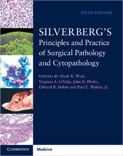
 Editors: Mark R. Wick, MD; Virginia A. LiVolsi, MD; John D. Pfeifer, MD, Edward B. Stelow, MD, and Paul E. Wakely, Jr., MD
Publisher: Cambridge University Press – 3,383 pages
Book Review by: Nano Khilnani
The term ‘pathology’ brings to mind the practice of examining tissue, bone, fluids and other components of the human body to determine the cause of death of the expired person. That is what autopsy pathologists typically do, Dr. Mack Wick points out in the first chapter.
He writes that surgical pathologists on the other hand look at specimens to examine conditions and diseases, then assist other physicians and surgeons to determine the best course of treatment for the patient. So the surgical pathologist is not only a researcher but also a consultant and teacher as well.
The surgical pathologist performs a critical role in the efforts of the entire hospital staff. He (or she) assists the team of health care professionals – doctors, medical residents, interns, and nurses – in their work to not only treat ill patients to make them well, but to also provide crucial information in order to save their lives. This important member of a medical center examines specimens received from various specialists and discusses his findings with them.
Among them are: dermatologists, endocrinologists, gastroenterologists, gynecologists, hepatologists, nephrologists, neurologists, oncologists, otolaryngologists, pediatricians, urologists, and physicians in other specialties.
Ninety-three experts in their respective fields – all from the United States, except one each from Australia and Canada – wrote the 48 chapters or contributed other material for the current edition of this four-volume book that has been in continuous print since 1983. Most of them teach pathology at various universities and/or practice it in hospitals and clinics.
Almost all -90- of the chapter authors are doctors of medicine, and 15 of those have in addition, PhD degrees in various fields of medical science. While most of the contributors have knowledge, training, and experience in pathology, some of them have backgrounds in these fields: dermatology, genetics, immunology, laboratory medicine, obstetrics-gynecology, oncology, ophthalmology, otolaryngology, and pediatrics.
This is an authoritative book with contributions from numerous respected physicians renowned in their specific areas. This is also a popular reference textbook in pathology as evidenced from its high ranking on the website of Amazon, the world’s largest online bookseller. Another reason why so many people use this book is that it has quite an extensive coverage of topics. We list below its chapters so that you the practitioner, medial resident, intern or student can get a view of what you will find covered in this book.
- Volume 1
- General philosophy and principles of surgical pathology and cytopathology, including quality and legal issues
- General and special techniques in surgical pathology and cytopathology
- Pathology of selected infectious diseases
- Immunological diseases and transplantation pathology
- The pathology of iatrogenic lesions
- Differential diagnosis of metastatic tumors
- Non-neoplastic skin disease
- Cutaneous tumors and tumor-like conditions
- Soft tissue tumors
- The breast
- Lymph nodes and spleen
- Bone marrow
- Volume 2
- Non-neoplastic diseases of bones and joints
- Neoplastic and tumor-like lesions of bone
- Sinonasal and nasopharyngeal pathology
- Larynx and trachea
- Non-neoplastic lung disease
- Tumors and tumor-like conditions of the lung
- Diseases of the serosal surfaces
- The cardiovascular system
- Surgical pathology and cytopathology of the thymus and mediastinum
- The oral cavity
- Diseases of the salivary gland
- Volume 3
- The esophagus
- The stomach
- Non-neoplastic diseases of the small and large intestines
- Neoplastic diseases of the small and large intestines
- Non-neoplastic diseases of the liver
- Neoplasms and tumor-like conditions of the liver
- The pancreas and extrahepatic biliary system
- Non-neoplastic renal diseases
- Neoplasms and tumor0like conditions of the kidney
- The urinary bladder, urachal remnants, urethra, renal pelves, and ureters
- The testis, paratesticular structures, and male external genitalia
- Diseases of the prostate
- Volume 4
- The uterine cervix
- The vulva and vagina
- The uterine corpus
- Ovary and fallopian tube
- The placenta, products of conception, and gestational trophoblastic disease
- The pituitary gland
- The thyroid gland
- The parathyroid glands
- The adrenal gland and tumors of extra-adrenal paraganglia
- Muscle and peripheral nerve pathology
- The ear
- The eye and ocular adnexa
- The central nervous system
This book comes with fully-searchable online access for purchasers, to all its text and images.
Here are the steps to set up your Cambridge Books Online account:
- Visit https://ebooks.cambridhe.org/register and click Register
- Enter your details and click Submit
- You will receive a confirmation email
- Follow the instructions in that email to activate your registration
Here’s how to activate your access code using your Cambridge Books Online account:
- Scratch off the tab on the inside front cover of your book to get your Activation Code
- Visit https://ebooks.cambridge.org and click Login, followed by User Login
- Log in using your Cambridge Books Online account
- Click on My Bookshelf
- Enter your Activation Code into the box provided and click Activate Now
- Click on the book title to view the book’s content
(This book cannot be returned once the access code has been revealed. For technical assistance go to: http://ebooks.cambridge.org/contact/jsf. Further information about using and navigating your online access may be found on the online User Guide once you have activated your code).
It is not only an immense and detailed reference work, it is also practice-oriented and user-friendly. It integrates the fields of surgical pathology and cytopathology by providing critically-needed information in a single source. For example, it presents relevant features of a particular lesion, side by side.
Some of the other important features of this book are the following:
- Over 3,500 color images depicting clinical features, morphological attributes, histochemical and immunohistochemical findings, and molecular characteristics of all lesions
- Editors who are highly experienced and academically accomplished
- Chapters written by leading experts in the field, with several new in this edition, bringing some fresh content
- Practical approach of Dr. Steven Silverberg carefully preserved in the present version of this text, providing trainees and practitioners with the relevant, seminal facts needed to make difficult but expeditious decisions in diagnosis and case management
- Pitfalls and interpretative snares are considered throughout
- Print book packaged with access to a secure electronic copy of the book, providing quick and easy access to its wealth of text and images
One of the most basic and important components of this book is chapter 2, General and special techniques in surgical pathology and cytopathology. This was authored by Drs. Mark R. Wick, Robert D. LeGallo, Edward B. Stelow, and John D. Pfeifer.
Let us take a look at what topics are presented and discussed in this chapter. It will give you an idea of how materials are organized and presented in other chapters of this book.
The chapter begins with an introductory couple of paragraphs that point out that there has been rapid growth and development of surgical pathology and cytopathology in the last few decades, with numerous conceptual and technical discoveries in basic sciences, particularly in biochemistry, cellular and molecular biology, and immunology.
The authors instruct the reader to proactively develop a plan of action to maximize the amount of information he or she can obtain from examination of a specimen. The purpose of the chapter is then briefly mentioned, which is primarily to highlight the techniques in surgical pathology and cytopathology.
Then, the topics and subtopics of chapter 2 are presented and discussed:
- Tissue fixation
- Histochemical staining methods
- Cytological samples and fine-needle aspiration biopsies (FNABs)
- Fine-needle aspiration and “core” biopsies, including fixations and stains for FNABs
- Electron microscopy
- Diagnostic immunohistochemistry
- Antigen (epitope) retrieval methods
- Background staining
- Interpretation of results and controls
- Immunohistology on cytological samples
- Regulatory issues in immunohistochemistry
- Automation and tissue microarrays in immunohistology
- Flow cytometry
- Basic principles of FCM and instrumentation
- Specimen disaggregation and preanalytic preparation in flow cytometry
- Isolation of nuclei or whole cells and staining methods
- Cellular quantity and flow cytometric analysis
- Ploidy assessment and histogram interpretation
- Coefficients of variation in flow cytometry
- S-phrase fraction
- Cell marker analysis
- Quality control in flow cytometry
- Laser scanning cytometry
- Image cytometry
- Basic principles of image cytometry and instrumentation
- Preanalytic preparation and image cytometry
- Image analysis
- Ploidy assessment
- Quantitative immunohistochemistry and image cytometry
- Morphometry
- Cytology and automation
- Cytogenetics
- Molecular biology
- Molecular techniques
- Southern blotting
- Restriction fragment length polymorphisms
- Polymerase chain reaction
- PCR variants
- Tissue in-situ hybridization
- ISH for measurement of mRNA
- ISH for analysis of chromosomes
- Chromogenic in-situ hybridization
- DNA sequence analysis
- Direct DNA sequence analysis
- Indirect DNA sequence analysis
- Next-generation sequencing
- Internet resources for DNA sequence analysis
- Microarray analysis
- Copy number and genotype analysis
- Gene expression analysis
- Microsatellite analysis
- Microsatellite instability assays
- Identity determination
- Epigenetic (methylation) analysis
- Analysis of clonality and loss of heterozygosity
- Molecular analysis of infectious diseases
- Bacteria
- Fungi
- Viruses
- Protozoans
The chapter ends with an Acknowledgment and a References list of 12 pages. This chapter, like others in the rest of this book contains several types of visuals that aid in the learning process. The most prevalent type of these visuals is stains, which are micrographs of specimens or biopsies.
You will also find numerous other types of graphics, such as anatomical drawings, charts, images taken through microscopes, photos of diseased skin and tissue, tables, and x-ray images. All these enhance learning, understanding, and retention.
In summary, this is an authoritative and comprehensive textbook on pathology in general, and surgical pathology and cytopathology in particular. It contains the combined sharing of the knowledge, experience, and insight of 93 experts in the field, most of whom are professors at medical schools and physicians in hospitals.
For a book that’s been in print for 32 years, there’s no better testimony to its worth and acceptance than that lengthy record itself. To say that it is an outstanding achievement is probably not sufficient. A more apt description is that it is a monumental contribution to medical knowledge and practice.
Editors:
Mark R. Wick, MD is Professor of Pathology in the Department of Pathology at the University of Virginia Health System in Charlottesville, Virginia.
Virginia A. LiVolsi, MD is Professor of Pathology and Laboratory Medicine in the Department of Pathology and Laboratory Medicine at the University of Pennsylvania Medical Center in Philadelphia, Pennsylvania.
John D. Pfeifer, MD, PhD is Vice Chair for Clinical Affairs, Pathology and Immunology; and Professor of Pathology and Immunology, and Professor Obstetrics and Gynecology at Washington University School of Medicine in St. Louis, Missouri.
Edward B. Stelow, MD is Associate Professor of Pathology in the Department of Pathology at the University of Virginia Health System in Charlottesville, Virginia.
Paul E. Wakely, Jr., MD is Professor of Pathology in the Department of Pathology at Ohio State University in Columbus, Ohio.
Technical Consultant:
James N. Lampros Jr., MD is with the Department of Pathology at the University of Virginia Health System.


