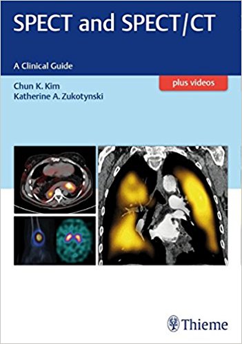
 Editors: Chun K. Kim, MD: and Katherine A. Zukotynski, MD
Publisher: Thieme – 205 pages, with 250 illustrations
Book Review by: Nano Khilnani
Single-photon emission computed tomography (SPECT), according to an article on Wikipedia, is “a nuclear medicine tomographic imaging technique using gamma rays. It is very similar to conventional nuclear medicine planar imaging using a gamma camera (that is, scintigraphy). However, it is able to provide true three-dimensional information. This information is typically presented as cross-sectional slices through the patient, but can be freely reformatted or manipulated as required.
“The technique requires delivery of a gamma-emitting radioisotope (a radionuclide) into the patient, normally through injection into the bloodstream. On occasion, the radioisotope is a simple soluble dissolved ion, such as an isotope of gallium. Most of the time, though, a marker radioisotope is attached to a specific ligand to create a radioligand, whose properties bind it to certain types of tissues. This marriage allows the combination of ligand and radiopharmaceutical to be carried and bound to a place of interest in the body, where the ligand concentration is seen by a gamma camera.”
Below is an animation of a SPECT scanning procedure on Wikipedia. Click on this link: https://en.wikipedia.org/wiki/Single-photon_emission_computed_tomography
SPECT/CT, the editors point out, is a combined or hybrid imaging procedure that yields the automated imaging of computed tomography (CT) with the functional capability of SPECT. These two imaging modalities are used in various medical specialties, and how they are used specifically in these areas is discussed in the chapters of this book listed below.
Seventeen specialists in cardiology, molecular imaging, nuclear medicine, neurology, radiology, radiochemistry, radiopharmacy, and therapeutics, from mainly the United States, but also two from Australia and one from Canada, authored the 11 chapters of this book, namely:
- The Fundamentals
- Basic Principles of SPECT and SPECT/Ct and Quality Control
- Radiopharmaceuticals for Clinical SPECT Studies
- Clinical Applications
- SPECT and SPECT/CT in Neuroscience
- SPECT and SPECT/CT for the Thyroid and Parathyroid Glands with Cases
- SPECT and SPECT/CT for the Cardiovascular System
- SPECT and SPECT/CT for the Respiratory System
- SPECT and SPECT/CT in Neoplastic Disease
- SPECT and SPECT/CT for the Skeletal System
- SPECT and SPECT/CT for Infection and Inflammation
- SPECT in Children
- Selected interesting SPECT and SPECT/CT Cases
To get the most out of the book – watch the videos at http://mediacenter.thieme.com When prompted during the registration process for your copy, enter the code found on the inside front cover of this book.
One medical specialty where these imaging techniques are used is cardiology. Their use is discussed in chapter 5, SPECT and SPECT/CT for the Cardiovascular System, pages 74-91. Let us take a look at the outline of this chapter and the topics and subtopics covered in it, to learn how content is laid out and discussed in other chapters of this book.
Chapter 5, SPECT and SPECT/CT for the Cardiovascular System, pages 74-91:
- Introduction
- Stress Testing and Myocardial Perfusion Imaging
- Stress Testing Protocols
- Myocardial Perfusion Imaging Protocols
- Troubleshooting Traditional and Novel SPECT MPI
- Fixed Anterior Wall Abnormalities
- Breast Attenuation
- Myocardial Scar and Hibernating Myocardium
- Fixed Inferior Wall Abnormalities
- Diaphragmatic Attenuation
- Excessive Subdiaphgramatic Activity
- Myocardial Infarction in the RCA Territory
- Reversible Perfusion Defects
- Patient Motion
- Misregistration
- Scanner Failure
- Balanced Ischemia
- Conclusion
- References
In addition, on each of the topics outlined above, the Pearls and Pitfalls are mentioned and discussed.
As technological innovations in imaging come to the fore, the process of diagnosing diseases becomes easier, with clearer and more detailed views of what is going on within a patient’s body.
This book on SPECT and SPECT/CT imaging is a unique one and helpful addition to the medical literature on diagnostic tools.
Editors:
Chun K. Kim, MD is Clinical Director of the Division of Nuclear Medicine and Molecular Imaging in the Department of Radiology at Brighma and Women’s Hospital, and Associate Professor of Radiology at Harvard Medical School in Boston, Massachusetts.
Katherine A. Zukotynski, MD is Associate Professor in the Departments of Radiology and Medicine at McMaster University in Hamilton, Ontario, Canada.
Here are interviews with various authors of Thieme books:


