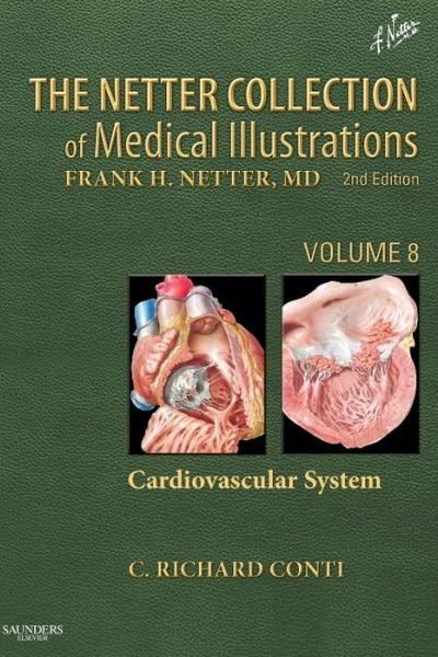
 Volume 8: Cardiovascular System
Editor: T. Richard Conti, MD. Additional Illustrations by: Carlos A.G. Machado, MD
Publisher: Elsevier Saunders – 295 pages
Book Review by: Nano Khilnani
Frank H. Netter, MD has been acknowledged worldwide by physicians and surgeons as the best medical illustrator who ever lived.
He was also one of the most prolific, as this book and eight others like it will clearly demonstrate to you. Each of the nine volumes in this series is focused on a particular system of the human body, and each contains numerous, detailed and anatomically precise drawings by Dr. Netter.
The nine books cover the reproductive, endocrine, respiratory, integumentary, urinary, musculoskeletal, digestive, nervous, and cardiovascular systems of the human body.
You will also find illustrations in this unique book by another renowned artist-physician Carlos A.G. Machado, a Brazilian cardiologist, who is the primary person responsible for continuing the Netter tradition of “painting masterpieces” and conveying “precise scientific information” as he aptly characterizes Dr. Netter’s work.
Four other contributing medical artists named at the end of this book review also have their illustrations in this particular volume on the cardiovascular system.
This book contains numerous full-color illustrations as well as detailed textual descriptions of the different parts of our cardiovascular system, namely the heart, and vessels going into and out of it. You will see the various types of cartilage, tendons, tissues and muscles within the heart, which itself has been described by many as a large muscle.
You will see the heart valves, pericardium, atria, auricles and ventricles as well as the aorta. You will see arteries (in red) along with veins (in blue), and various nerves.
There are also drawings of other organs next to the heart such as the lungs (and their parts), bronchial tubes, the diaphragm, the esophagus, the larynx and pharynx, the ribs, spine, sternum, trachea, thymus, and other adjacent organs and blood vessels.
It is organized into six sections, namely:
- Anatomy
- Physiology
- Imaging
- Embryology
- Congenital Heart Disease
- Acquired Heart Disease
Numerous study tools such as black-and-white and color charts, computer tomography (CT) scans, echocardiograms, magnetic resonance images (MRIs), photos, radiographs, tables and x-rays abound in the book, hastening your learning, understanding and retention.
The student and practitioner will find hundreds of “works of original art” by Drs. Netter and Machado and others. These are parts of the human heart and adjacent organs and systems representing accurate, exact and up-to-date medical knowledge.
Dr. Conti has done an expert job of choosing and presenting the pictures and other material, along with the help of Dr. Machado and the other medical illustrators named below. He has also written the text of this entire book – a monumental achievement. He deserves much credit.
Editor: C. Richard Conti, MD, MACC, FESC, FAHA is Emeritus Professor of Medicine at the University of Florida College of Medicine in Gainesville, Florida.
Associate Medical Illustrator: Carlos A.G. Machado, MD, who provided other illustrations for this book.
Contributing Illustrators:
Tiffany S. DaVanzo, MA, CMI
John A. Craig, MD
James A. Perkins, MS, MFA
Anita Impagliazzo, MA, CMI
Advisory Board:
A John Camm, QHP, MD
Larry Cochard, PhD
J. Michael Criley, MD
Anthony DeMaria, MD
Eugenio Gaudio, MD
Hyo-SooKim, MD, PhD
Bruce T. Liang, MD
Robert Roberts, MD, MACC


