 Author: Grace C.H. Yang, MD
Publisher: Cambridge University Press – 462 pages, with 2,000+ photomicrographs in CD
Book Review by: Nano Khilnani
Fine-needle aspiration (FNA) is a method to diagnose disease in lumps or masses of tissue. In this procedure, a thin (such as 23-25 gauge) hollow needle is inserted into the mass for sampling of cells that, after being stained, will be examined (biopsied) under a microscope to check if there is, or isn’t evidence of disease, or something unusual or unexpected..
The sampling and biopsy considered together are called fine-needle aspiration biopsy (FNAB) or fine-needle aspiration cytology (FNAC). The latter procedure refers to cell examinations only.
Fine-needle aspiration biopsies are very safe, minor surgical procedures. Often, a major surgical procedure – such as an excisional or open biopsy – can be avoided by performing a needle aspiration biopsy instead.
In 1981, the first fine-needle aspiration biopsy in the United States was done at Maimonides Medical Center in Brooklyn, New York, eliminating the need for surgery and hospitalization. Today, this procedure is widely used in the diagnosis of cancer and inflammatory conditions.
A needle aspiration biopsy is safer and less traumatic than an open surgical biopsy, and significant complications are usually rare, depending on the body site. Common complications include bruising and soreness.
There is a risk of loss of time if nothing wrong is found, if the biopsy is very small involving only a few cells. Problematic cells can be missed, resulting in a false negative result, or that the cells taken will not enable a definitive diagnosis.
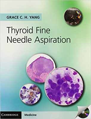
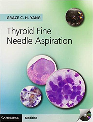
This 2013 book of over 460 pages, completely authored and edited by Dr. Grace Yang, is on fine-needle aspiration diagnostic techniques involving thyroid lesions and tumors, both of a cancerous and benign nature. She has performed over 10,000 ultrasound-guided thyroid fine-needle aspirations, and is highly knowledgeable in this specialty within both medicine and science.
It consists of 16 chapters supplemented with case histories and illustrations found in two appendices. We list below the titles of its chapters to provide you a broad overview of its coverage:
- Techniques and approaches to ultrasound-guided fine needle aspiration
- Nodular goiter and mimickers
- Follicular neoplasm and mimickerts
- Hurthle cell neoplasm and mimickers
- Follicular variant of papillary carcinoma
- Thyroiditis and mimickers
- Papillary thyroid carcinoma and variants with papillae and mimicker
- Papillary thyroid carcinoma, uncommon variants, and mimicker
- Clear cell and mucinous thyroid tumors
- Poorly differentiated thyroid carcinoma
- Anaplastic thyroid carcinoma
- Medullary thyroid carcinoma
- Miscellaneous benign lesions
- Metastastic thyroid carcinoma
- Metastastic and secondary tumors of the thyroid
- Ancillary tests
- Index to case histories
- Index to ultrasound and Doppler images
- General index
This book comes with a Compact Disk (CD) attached on its inside back cover.
This volume provides concise discussions and imaging of cases from 350 patients. A large range of thyroid lesions are shown in it, enabling readers see and understand their complexity and variety, and help them in their task of diagnosis. These clinical presentations correlate cytology and histology, as well as pathology and radiology.
Similar-looking lesions are shown together, so comparisons of differences can be done more easily. Dr.Yang also points out mistakes that are made in diagnoses, and their adverse and negative consequences. She shows students and clinicians how to avoid such pitfalls.
Let us take a look at the outline of topics and some images found in chapter 14 entitled Metastastic Thyroid Carcinoma on pages 422-440:
- Lymphatic versus hematogenous metastasis
- Different morphology between primary and metastastic thyroid carcinoma
- Similarity between cystic variant of papillary carcinoma and cystic lymph node metastasis of papillary carcinoma
- Detection of thyroglobulin in needle rinses of nonthyroidal neck masses
- Clues from ultrasound
- Lateral aberrant thyroid
References
A large number of micrographic images (about 130, including small insets within larger images) of various sizes are provided in the 20 or so pages of this chapter. These provide useful comparisons and differences in the various conditions, diseases and disorders shown. Detailed photo captions accompanying these images help the reader understand what has been going on in the cells and tissues of the patients.
This is a highly valuable resource for healthcare professionals who examine and treat patients with various types of lesions. It is useful for medical students, residents, and physicians. It is a particularly important book to have in the medical collections and libraries of cytopathologists, endocrinologists, histologists, histopathologists, radiologists, sonographers, surgical pathologists, and of course, surgeons.
Author and Editor:
Grace C.H. Yang, MD is Clinical Professor of Pathology and Laboratory Medicine at Weill Medical College on Cornell, New York.


