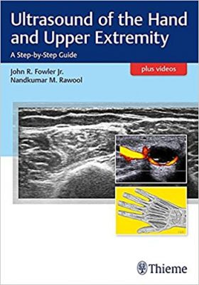
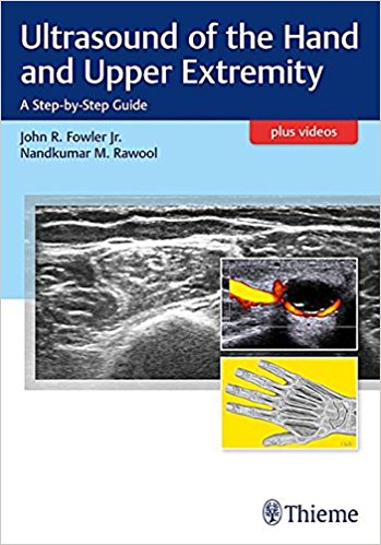
 Editors: John R. Fowler, Jr. MD; and Nandkumar M. Rawool, MD
Illustrator: Andrea Hines
Publisher: Thieme – 188 pages
Book Review by: Nano Khilnani
This compact (7”x10”) book with online videos (see link below) is ideal for busy clinicians, radiologists and hand surgeons. It is a practical, handy guide that serves as a quick reference source, with bullet-points and full-color and back-and-white illustrations: charts, fine-line drawings, photos, sketches, and ultrasound images.
Thirteen specialists in radiology, as well as in orthopedics, physical medicine, and plastic surgery from Florida, Texas, and Pennsylvania, including seven affiliated with the University of Pittsburgh, authored the 16 chapters of this book listed below:
- Section I. Introduction
- Basic Tenets of Upper Extremity Ultrasound
- Image Optimization in Musculoskeletal Ultrasound Imaging
- Section II. Fingers and Wrist
- Trigger Finger
- Evaluation of Flexor Tendon Lacerations
- Carpal Tunnel Evaluation and Injection
- First Dorsal Compartment Tendonitis
- Evaluation of Wrist Joints
- Section III. Forearm and Elbow
- Evaluation of Nerves of the Elbow and Forearm
- Evaluation of Lateral Epicondylitis
- Evaluation of Collateral Ligaments
- Evaluation of Distal Biceps
- Section IV. Shoulder
- Shoulder Examination
- Ultrasound Guided Shoulder Injections
- Treatment of Adhesive Capsulitis
- Section V. Masses
- Evaluation of Hand and Finger Masses
- Appearance of Common Masses
Accompanying Videos:
- 3.1: Identification of the A1 pulley
- 5.1: Anisotropy of the median curve
- 5.2: Flexor pollicis longus contraction of the median nerve
- 5.3: Median nerve from the forearm to the carpal tunnel
- 6.1: deQuervain ultrasound
- 8.2: Median nerve
- 8.3: Radial nerve
- 9.1: Lateral epondylitis ultrasound
- 10.1: Dynamic evaluation of the ulnar collateral ligament
- 11.1: Dynamic evaluation of the distal biceps using a lateral approach
- 11.2: Dynamic evaluation of the distal biceps using a medial approach
- 11.3: Dynamic evaluation of the distal biceps using a posterior approach
To watch these videos, retrieve the access code on the first page of this book and register your copy at www.MediaCenter.Thieme.com
Like all imaging modalities, ultrasound has its plusses as well as its minuses. Here they are:
Advantages of Ultrasound:
- Non-ionizing sound waves
- Readily available
- Portable
- Inexpensive
- Real-time evaluation of a specific problem area
- Allows rapid assessment of multiple joints for side-to-side comparison
- Useful for evaluation of soft tissue structures, including ligaments and nerves
- Allows for evaluation of structures under tension and with motion
- Confirms appropriate needle position for joint capsule injections
Disadvantages of Ultrasound:
- Extremely operator dependent
- May fail to define the full scope of a presenting problem with problem-focused scanning
- Cannot evaluate tissues with high acoustic impedance (bone and air)
- Risk of thermal heating or mechanical injury with high frequency
- Many artifacts
- Limited value in obese and morbidly obese patients
- Limited visualization of structures deep to the cortical surface of bone
- Limited visualization of intra-articular structures
- Time-consuming to complete an appropriate examination
Some of the major features of this book are:
- Full-color photographs to depict proper patient and probe positioning for optimal effects
- Expert advice on ultrasound machine settings for achieving the best images in various structures
- Labeled ultrasound images of deformities and normal anatomy for comparative clinical use
- Thirteen instructive videos highlight ultrasound techniques for a range of structures and pathologies
This is a compact, excellent, practical, and easy-to-use guide for residents and practicing radiologists and surgeons of the hand and upper extremity.
Editors:
John R. Fowler, Jr. MD is Assistant Professor in the Department of Orthopedic Surgery, and Assistant Dean of Medical Student Research at the University of Pittsburgh Medical Center in Pittsburgh, Pennsylvania.
Nandkumar M. Rawool, MD, RDMS is Associate Professor in the Department of Radiologic Sciences, and Program Director of Diagnostic Medical Sonography and Cardiovascular Sonography at Thomas Jefferson University in Philadelphia, Pennsylvania,
Illustrator:
Andrea Hines


