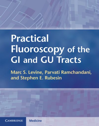
 Authors-Editors: Marc S. Levine, MD; Parvati Ramchandani, MD; Stephen E. Rubesin, MD
Publisher: Cambridge University Press – 224 pages
Book Review by: Dev Daulat
The use of cross-sectional imaging means such as computed tomography (CT), magnetic resonance (MR), and positron emission tomography (PET) has been on the rise in recent years, but radiology practitioners continue to perform gastrointestinal (GI) and genitourinary (GU) fluoroscopic procedures on a daily basis, the editors point out at the outset of this book, in the Preface.
They point out that it is “important for radiologists to develop and maintain their skills in GI and GU fluoroscopy in order to provide optimal studies for their patients,” and for this reason the three of them got together to produce this book “dedicated to all aspects of performing and interpreting high-quality GI and GU fluoroscopic procedures.”
I was looking for an explanation in the Preface as to why they prefer to use fluoroscopy procedures rather than the other three imaging modes they mentioned, or some sort of summary of the differences between the CT, MR, and PET scan methods as a group on the one hand, and fluoroscopy on the other. I did not find any such “compare-and-contrast” passage.
They do state however that fluoroscopy is critical in some ways: “Our goal in writing this text was to illuminate the critical role of fluoroscopy in the diagnosis of luminal GI and GU diseases.”
This book presents the “fundamentals and nuances” of performing fluoroscopy, as well as contains numerous fluoroscopic images of organs and surrounding areas within the GI and GU tracts, along with detailed interpretations of the findings in each instance. The areas of coverage are listed below to give you – the practicing radiologist, assistant, or trainee – an overview of what you will find in this book:
- Section I – GI Tract
- Pharynx
- Examination of the esophagus, stomach, and duodenum: techniques and normal anatomy
- Esophagus
- Stomach
- Duodenum
- Examination of the small intestine: techniques and normal anatomy
- Small intestine
- Examination of the colon: techniques and normal anatomy
- Colon
- Section II – GU Tract
- Fluoroscopic evaluation of the bladder, urethra, and urinary diversions
- Retrograde pyelography
Among the types of images and interpretive discussions of the findings you will discover in the GI section of this book are the following:
- All types of postoperative GI studies
- Double- and single-contrast barium enemas
- Double- and single-contrast esophagography
- Double and single-contrast upper GI exams
- Enteroclysis
- Evacuation proctography
- Modified barium swallow
- Videofluoroscopic pharyngography
In the GU section are chapters on the following fluoroscopic imaging modalities of particular anatomies:
- Retrograde pyelourethrography
- Retrograde urethrography
- Voiding cystography
The “how-to’s” of the techniques of conducting various fluoroscopic procedures are laid in this unique text, along with advice based on experience and insight of the authors, solutions to pitfalls and problems, and valuable tips.
This book also contains:
- Discussions on important clinical and radiographic findings
- Annotated images to illustrate these findings
- Relevant differential diagnoses for the major pathological conditions
The organization and layout of content makes it easy for you to understand, retain and recall information. Let us take a look below at chapter 2, Examination of the esophagus, stomach, and duodenum: techniques and normal anatomy, in terms of these important elements. Note that there are various subtopics within the sections below that we have not shown below. Also, keep in mind that relevant images are presented in each section, with informational captions.
- Introductory paragraph
- General principles
- Materials
- Routine techniques
- Variations in technique: problems and pitfalls
- Normal anatomy and anatomic variations
- Gastric outlet obstruction
- Gastric resection
- References
All in all, this is a great book in its field.
Editors:
Marc S. Levine, MD is Chief of the Gastrointestinal Radiology Section at University of Pennsylvania Medical Center, and Advisory Dean at Perelman School of Medicine at the University of Pennsylvania in Philadelphia, Pennsylvania.
Parvati Ramchandani, MD is Chief of the Genitourinary Section at University of Pennsylvania Medical Center; and Professor of Radiology and Surgery at Perelman School of Medicine at the University of Pennsylvania in Philadelphia, Pennsylvania.
Stephen E. Rubesin, MD is affiliated with the Gastrointestinal Radiology Section at University of Pennsylvania Medical Center, and is Professor of Radiology at Perelman School of Medicine at the University of Pennsylvania in Philadelphia, Pennsylvania.


