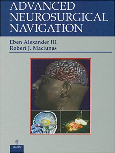
 Editors: Eben Alexander III, MD; and Robert J. Maciunas, MD
Publisher: Thieme – 605 pages
Book Review by: Nano Khilnani
Detailed imaging of the human nervous system is a critical imperative in successful neurosurgery, the two editors point out at the outset of this large book, in their Preface.. “The complex functional anatomy intricately woven into the central nervous system is often distorted by local pathological processes as well as by the ‘rewiring’ of the healing brain,” they write, as they explain the difficulties in neurosurgery.
Technological advancements have certainly helped neurosurgeons better visualize the complex structural system in our brains. One of these advances is stereotaxy, a three-dimensional viewing platform used not only in neurosurgery but also in operations in other parts of our bodies. Examples of some of the stereotactic images can be seen in pages 115-123.
Eben Alexander III and Blaine S. Nashold, Jr. write as authors of chapter 1 – A History of Neurosurgical Navigation – that at the core of the stereotactic methodology lays the construction of a stereotactic frame. This frame is based on a series of rectilinear coordinates for determining a specific target point in the human brain.
Designed by the French philosopher and mathematician Rene Descartes in the seventeenth century, he stated that any point in three-dimensional space can be defined by its relationship with three intersecting planes at 90 degrees to each other. These planes are designated as x, y, and z coordinates and were oriented at right angles to each other.
In the stereotactic arc system, the authors of the chapter explain that the target is localized at the center of a semicircular arc. Several techniques have been used:
In one, the stereotactic instrument is placed directly on the skull, and the arc allows the positioning of the probe or electrode to be introduced to the center of the arc, which is the target point.
A second method allows the use of a phantom base and target point. The electrode is prepositioned on the phantom and then transferred to the fixed frame on the patient’s head (Reichert-Wolf instrument). The accuracy of these instruments is based on their precise construction.
Attachment of the frame to the patient’s head can be done with four techniques, each one explained in the aforementioned chapter.
Stereotactic neuro-imaging technology enables advanced neurosurgical navigation, the subject of this book. One of the important values then, of this volume published in 1999 is the multi-dimensional images shown throughout over 600 pages.
Technology holds further promise, the editors note optimistically. They write: “Today, neurological surgery stands at a technological crossroads. The last decade has witnessed unprecedented advances in neurosurgical techniques. Revolutionary advancers in high-speed graphic computers, informatics, biotechnology and robotics promise to metamorphose the practice of this therapeutic discipline. The very boundaries and definition of the field are subject to radical transformation as a result of these fundamental changes.” In short, excitement abounds in neurosurgery.
One hundred and twenty-one specialists in neurology, neurosurgery and other fields such as artificial intelligence, bioinformatics, biophysics, computer science, pharmacology, physiology and radiation oncology – from all over the United States and seven other countries – Belgium, Canada, Egypt, France, Sweden, Singapore and the United Kingdom – wrote the 48 chapters of this wide-ranging book.
The content is organized around five Parts we list below that provides you a broad overview of the scope of this unique text.
- Part I. History and Overview
- Part II. Transferring Medical Data into Maps
- Part III. Stereotaxy: Clinical Applications
- Part IV. Image-Guidance for Epilepsy, Radiation and Functional Neurosurgery
- Part V. Frontiers in Neurosurgical Navigation
Let’s take a close look at the contents of chapter 21 – Stereotactic Biopsy of the Brain Stem and Posterior Fossa – found on pages 279-300, and chapter 22 – Steretactic Frame-Based Guidance of Craniotomies: Cosman-Roberts-Welsl System. Here you can see and understand how stereotactic imaging techniques work.
In chapter 21 – Stereotactic Biopsy of the Brain Stem and Posterior Fossa, after a brief introduction, the topics discussed are:
- Stereotactic Techniques
- Stereotactic Basics
- University of Florida Methods
- Trans-frontal Approach
- Trans-cerebullar Approach
- Trans-tentorial Approach
- Indications
- University of Florida Experience
- Discussion
- Complications: Prevention and Management
- Conclusions
- References
In chapter 22 – Steretactic Frame-Based Guidance of Craniotomies: Cosman-Roberts-Welsl System – after a brief two-paragraph introduction the topics covered are:
- Materials and Methods
- Frame Application
- Database Acquisition and Treatment Planning
- Surgical Procedure: Resection
- Surgical Procedure: Transplantation
- Clinical Material
- Presenting Symptoms
- Neurodiagnostic Imaging (Vanderbilt University Hospital Series)
- Histology
- Results
- Operative Time
- Target Localization
- Intra-operative Ultrasonography
- Postoperative Computed Tomography / Magnetic Resonance Imaging Scanning
- Correlation of Imaging and Histology
- Postoperative Status
- Transplantation Results
- Infections
- Postoperative Survival
- Multiple Recurrent Lesions (Vanderbilt University Hospital Series)
- Discussion
- Conclusions
- References
This uniquely important book is a very useful, practical and detailed one in navigating the brain accurately and precisely, which is critical to successful outcomes in neurosurgery.
Editors:
Eben Alexander III, MD is Associate Professor of Surgery and Neurosurgery and Assistant Professor in Radiation Therapy in the Department of Neurosurgery at the Brigham and Women’s Hospital. He is also affiliated with The Children’s Hospital, the Massachusetts, General Hospital, and at Harvard Medical School in Boston, Massachusetts.
Robert J. Maciunas, MD is Frank P. Smith Professor and Chairman of Neurological Surgery at the University of Rochester Medical Center in Rochester, New York.


