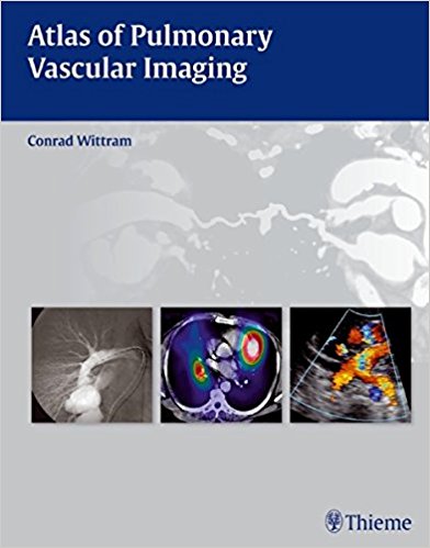
 Author: Conrad Wittram, MB
Publisher: Thieme – 161 pages
Book Review by: Nano Khilnani
Imaging technologies have continued to improve over the years, especially computed tomography (CT) and magnetic resonance imaging (MRI), Dr. Lawrence R. Goodman points out in his Foreword to this book. He is Professor and Director of Diagnostic Radiology and Pulmonary Medicine, and Director of Thoracic Imaging at the Medical College of Wisconsin in Milwaukee.
These days, better spatial resolution, contrast resolution, and temporal resolution enable the images of pulmonary vessels in their relation to other intrathoracic structures, to be seen in “exquisite vascular detail,” something that was never possible before, he points out. Such detailed imaging has enhanced the diagnosis of many congenital and acquired diseases, and improved understanding of the underlying pathophysiology.
Dr. Conrad Wittram, the author of this concise volume on the imaging of vessels in the lung, also has praise for present-day enhanced technologies: “Multidetector computed tomography has revolutionized the imaging of acute pulmonary embolism and ushered in a new radiology discipline: pulmonary vascular imaging.
This book provides a comprehensive and systematic approach to illustrate common, uncommon, and quite rare pulmonary conditions and diseases through these imaging modalities:
- Angiography
- Computed tomography
- Magnetic resonance imaging
- Nuclear medicine
- Radiography
- Ultrasound
It has been written for use by cardiologists and cardiothoracic surgeons, pulmonologists, and radiologists, and medical residents and fellows in these specialties. The author writes that it “provides a foundation of understanding and encourages interest and further research in pulmonary vascular disease.” Here are the 12 chapters of this relatively concise volume:
- Anatomy
- Congenital Anomalies
- Cardiac Disease
- Embolism
- In Situ Thrombosis
- Aneurysm and Varix
- Vasculitis
- Infection
- Trauma and Intervention
- Tumor
- Systemic and Lung Diseases
- Pulmonary Arterial Hypertension
The chief values of this book that make it an outstandingly unique and easy to use are that it provides a large number of images and a bullet-point format. Detailed captions are provided for each image presented, enabling the reader to understand the pathophysiology of each abnormality, anomaly, condition, disease, or disorder presented.
Let us take a look at chapter 2, Congenital Anomalies. After a brief introductory paragraph about inherited anomalies in pulmonary arteries and veins found at birth or later in life, the chapter presents and discusses in detail, though images and bullet points, the various known anomalies:
- Pulmonary Artery Agenesis
- Pulmonary Artery Hypoplasia
- Pulmonary Artery Stenosis
- Left Pulmonary Artery Sling
- Truncus Arteriosus
- Pulmonary Arteriovenous Malformation
- Systemic Artery to Normal Lung
- Intralobar Sequestration
- Extralobar Sequestration
- Hypogenetic Lung (Scimitar) Syndrome
- Bronchial Arteriovenous Malformation
- Partial Anomalous Pulmonary Venous Return
- Total Anomalous Pulmonary Venous Return
A list of Suggested Reading materials is provided at the end of this chapter.
Among the key benefits of owning this book are the following:
- In-depth coverage of how the pulmonary vessels are affected by congenital anomalies, cardiac disease, emboli, in situ thrombosis, vasculitis, tumors, aneurysms, and other lung vessel pathologies
- 322 high-resolution images demonstrating a wide variety of imaging modalities, from radiography, angiography, and multi-slice CT, to MRI, ultrasound, and nuclear imaging
- Succinct bullet-point format enabling quick and easy reference
- High-quality Angiographic and correlative CT images and instructive drawings illustrating the diagnostic criteria of pulmonary embolism
- Tips on how to recognize pulmonary embolism mimics, such as partial volume and flow-related artifacts
Author:
Conrad Wittram, MB, ChB is Associate Professor in the Department of Radiology at Harvard Medical School and at Massachusetts General Hospital in Boston, Massachusetts.