 Authors: Elizabeth A. Montgomery and Lysandra Voltaggio
Publisher: Wolters Kluwer | Lippincott Williams & Wilkins – 328 pages
Book Review by: Nano Khilnani
This book is for at least two types of medical specialists. The first is gastroenterologists or those specialize in curing diseases of the gastrointestinal (GI) tract which includes the esophagus, stomach, small intestine, large intestine or colon, and the rectum. In some medical textbooks, such as the present one, the anus is sometimes considered the last part of the GI tract, instead of the rectum.
The other type of specialists are pathologists, or those who study the causes of disease or abnormalities in some parts of the body, or deviations in structure and / or functions from what is considered normal.
This volume is limited to the study of neoplastic mucosal biopsies. As most people know, a biopsy is the removal of cells, fluids and tissues from the human body for the purpose of examination. The word ‘mucosal’ refers to the mucous membrane lining the inside walls of parts of the GI tract. But most people may not know what neoplastic means. The word means ‘tumorous’ or, relating to, or constituting a tumor, coming from the word neoplasia which is either the process of the formation of tumors, or a tumorous condition.
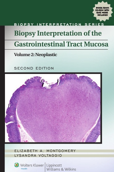
This small-size (6” x 9”) book of over 300 pages presents some 280 four-color images and descriptions of biopsy samples taken from the five parts of the GI tract we name above which are the esophagus, stomach, small intestine, colon, and anus. The photos are on average a half or a third the size of the book’s pages.
Besides these photos in the printed book, an additional around 1400 images are found in the online version of this book, which can be found on this web address with an access code: http://solution.lww.com/BISGITractMucosa2eV2Neoplastic. The access code is available to purchasers of this book by scratching off a label found on the inside front cover of this book.
Elizabeth A. Montgomery and Lysandra Voltaggio, the authors of this work, write that this book is “intended as a practical reference for pathologists to use when making diagnostic decisions based on biopsy specimens…this is a concise and comprehensive volume covering the interpretation of neoplastic GI mucosal biopsies. Content is geared to daily practice and includes mucosal biopsies…” (from the parts of the GI tract we named earlier).
The authors, writing in their Introduction about the general features of interpreting biopsies, point out that with respect to a host of injuries, there is a very limited number of responses available within the GI tract. So diagnosing the type of injury in a given biopsy requires correlation with information available from an endoscopy (examination of a number of internal organs) or other clinical procedure.
They also caution that the pathologist not succumb to merely reporting “inflammation” when examining specimens. This is because inflammatory cells are a normal part of the lamina propia. The lamina propia is vascular layer of connective tissue under the basement membrane. The basement membrane is a layer of the epithelium or topmost part of the internal wall of parts of the GI.
In order to understand all this and other material in this book, we suggest that readers go online and actually see the internal structures and functions of the organs that constitute the GI tract.
Elizabeth A. Montgomery, MD is affiliated with the department of pathology at the Johns Hopkins Medical Institutions in Baltimore, Maryland.
Lysandra Voltaggio, MD is associated with the department of pathology at George Washington University Hospital in Washington, D.C.