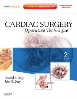
 Authors: Donald B. Doty, MD and John R. Doty, MD. Illustrations by Jill Rhead, MA, CMI, FAMI and Christy Krames, MA. CMI, FAMI
Publisher: Elsevier Saunders – 630 pages
Book Review by: Nano Khilnani
Manufactured with superior-quality glossy paper, this 9” x 11” hardcover book, the publisher has enabled presentation of clear, detailed, full-color anatomical illustrations and photos of various organs in the cardiovascular system on every right side page of this entire book except in the Index section at the end. This is an excellent way to learn cardiovascular surgery: read the text on the left while you’re looking on the right what has been written about.
For example, let us take a look at chapter 1 entitled Cardiac Anatomy, which starts out on the left page (page 2, with page 1 being the chapter title page) with these words:
“Understanding cardiac anatomy is fundamental to success in cardiac surgery. The following drawings show the cardiac surgeon’s view of the heart from the right side of the supine patient through a mid-sternal incision. Photographs illustrate the anatomic position as if looking at an upright patient, unless otherwise noted.”
Below the above block of text are four other blocks of text labeled A, B, C, and D under the heading Figure 1-1. On the right-side page are four views of the heart from different positions.
Figure A shows one view of the heart and the text on it on the left reads: “The cardiac structures most accessible to view are the superior vena cava, right atrium, right ventricle, main pulmonary artery, and aorta. Only a small portion of the anterior wall of the left ventricle is visible. Medial displacement of the right atrium exposes the left atrium and right pulmonary veins.”
Figure A is labeled with names of the various visible structures of the heart, clockwise: pulmonary artery, right ventricle, inferior vena cava, right pulmonary veins, right pulmonary artery, superior vena cava, aorta, and left pulmonary artery.
Chapter 31 entitled Aortic Valve Replacement is 52 pages long, and explains on the left-sided pages and illustrates on the right-sided pages with drawings and actual full-color photos of surgical procedures, the steps that are taken in this type of surgery, to accomplish the various tasks in the replacement of this important part of the heart.
The contents in this book are presented in 45 chapters grouped together in 12 Parts, namely:
- Basic Considerations
- Septal Defects
- Anomalies of Pulmonary Venous Connection
- Right Heart Valve Lesions (Congenital)
- Left Heart Valve Lesions (Congenital)
- Single Ventricle
- Malposition of the Great Arteries
- Thoracic Arteries and Veins (Congenital)
- Valve Lesions (Acquired)
- Ischemic Heart Disease
- Thoracic Arteries and Veins (Acquired)
- Special Operations
Included for you as the purchaser of this print edition of the book is a very user-friendly accompanying website www.ExpertConsult.com which is an online interactive learning platform that presents a vast collection of Elsevier textbook titles with a wide variety of ancillary material.
The website features:
- Fully searchable text
- Integration links that will seamlessly connect you to additional and related content in other ExpertConsult titles
- An image library, with figures that can be easily downloaded into PowerPoint
- Supplementary material such as audio or video clips
If you are already a registered user at ExpertConsult, you can gain access to the online version of this book by going to the above-mentioned website and entering the unique PIN code provided in the scratch-off box on the inside front cover of this book.
If you are a first-time user, do the following:
Register
- Click on Register Now at www.expertconsult.com
- Fill in your user information and click Continue Activate
Activate your book
- Scratch off your Activation Code in the inside front cover of your book
- Enter it into the Enter Activation Code box
- Click Activate Now
- Click the title under My Titles
For technical assistance you can email: [email protected], or call: 800-401-9962 inside the U.S. or 1-314-995-3200 outside the U.S.
There’s probably no easier way to a acquire detailed anatomic knowledge of the heart and learn about a wide range of cardiac surgical procedures than through a book, and this one is one of the best I have come across so far, with credit for it going to the following:
Authors:
Donald B. Doty, MD and John R. Doty, MD are with the Division of Cardiovascular and Thoracic Surgery at the Intermountain Medical Center in Salt Lake City, Utah.
Medial Illustrators:
Jill Rhead, MA, CMI, FAMI of Salt Lake City Utah.
Christy Krames, MA, CMI, FAMI of Austin, Texas


