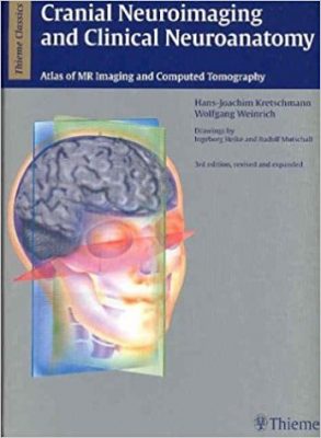
 Authors: Hans-Joachim Kretshmann, MD; and Wolfgang Weinrich, MD
Drawings by: Ingeborg Heike and Rudolf Mutschall
Publisher: Thieme – 451 pages, with 664 illustrations
Book Review by: Nano Khilnani
This large-size (9”x12”) atlas of the various networks of nerves provides an average of nearly one and a half images per page, and with the Index excluded, it works out to much more than one and a half images per page. Some pages have as many as four (smaller) images, with detailed notes correlating to numbered points marked on the images.
According to the authors, the content in all its chapters has been updated in this third English edition published in 2004. The book also has editions in German and Japanese languages, something that is quite rare in medical book publishing.
That this book is now in its third edition is a testament to its academic and commercial success. The first edition released in 1984 was quite well received, and the discovery of new knowledge about the brain necessitated an updated second edition in 1992. This current edition has the following new features and updates:
- The number of images have almost doubled
- Old CT and MR images have been completely replaced with large-sized illustrations
- Additional images include those on vascular supply to the posterior fossa
- Arterial territories of the infratentorial space are introduced in this edition
- Line drawings taken from anatomic specimens provide a clearer picture of the anatomy than the actual MR images, each of which is then outlined and highlighted to identify the various anatomic structures that are then also annotated in great detail
- Clear, color-coded identification of vascular supply to various areas of the brain on individual slices have been added. This is very helpful in today’s practice where early stroke detection and localization of the involved areas are vital prior to decisions on interventional procedures
This book has been designed for everyday practice to enable physicians to determine if the symptoms the patients describe correlate with the findings revealed by the images. The intended users are neuroanatomists, neurologists, neurophysiologists, neurosurgeons, oncologists, and traumatologists, as well as medical students, residents, and fellows.
Below are the titles of the chapters in this third edition. Within the book is a more detailed listing of the topics covered and discussed in each of the chapters, along with lots of images accompanying the discussions:
- Introduction
- Neuroimaging and Guideline Structures
- Topography of the Facial Skeleton and the Cavities in Multiplanar Parallel Slices
- Topography of the Pharynx and Craniocervical Junction in Multiplanar Parallel Slices
- Topography of the Neurocranium and Its Intracranial Spaces and Structures in Multiplanar Parallel Slices
- Neurofunctional Systems
- Neurotransmitters and Neuromodulators
The importance for specialists in neurology, especially neurosurgeons, to have a three-dimensional knowledge of anatomic structures cannot be overemphasized when the reader browses through the chapters of this book. Let us take a look at how this point is illustrated and discussed in chapter 3 – Topography of the Facial Skeleton and its Cavities in Multiplanar Parallel Slices, on pages 210-229.
On the first page of this chapter, the authors state: “The exact position of the slices is important in neuroanatomy,” then refer to line drawings on subsequent pages. The various areas of the facial anatomy are marked with different colors. The areas shown are: bones and muscles of the craniocervical junction, nasal cavity and paranasal sinuses, parapharyngeal space, oral cavity, orbit, and the pharynx.
When the reader views these images, he realizes that it is critical for the neurosurgeon to know what lies not only on the left and right sides of the anatomic part he is working on, but also what is on the bottom, and as a matter of fact, what are all around where he is performing surgery. This chapter, as is true of all chapters of this book, provides detailed fine-line drawings. These drawings need to not only be studied, but also committed to memory, if the surgeon is to avoid mishaps in surgery.
Painstaking work in making this a detailed atlas has no doubt resulted in very useful features:
- Anatomic terminology that has been updated to international standards
- Broad range of images representing brain structures, arteries, arterial territories, nerves, veins, and neurofunctional systems
- Expanded and updated timely information on this highly complex specialty
- High-resolution T1- and T2-weighted MR images making localization simpler and more efficient
- More than 640 meticulously detailed illustrations of all the important cranial computed tomography (CT) and magnetic resonance (MR) images in three planes: axial, coronal and sagittal.
This is an essential reference work to have, for all specialists in neurology, neurosurgery and allied fields. It provides detailed views of our complex brain.
Editors:
Hans-Joachim Kretshmann. MD is professor and former director of the Department of Neuroanatomy at the Hanover Medical School in Hanover, Germany.
Wolfgang Weinrich, MD is professor and former head of the Neurological Clinic at Nordstadt Hospital in Hanover, Germany.