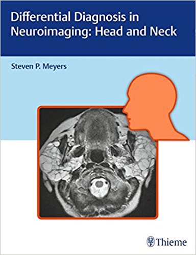
 Author: Steven P. Meyers, MD
Publisher: Thieme – 649 pages, with 1,538 illustrations
Book Review by: Nano Khilnani
This book is based on Dr. Steven P. Meyers’ three decades of collection of notes and data including 25 years working as an academic neuroroadiologist at a university medical center.
He has used them as a teaching file for lectures and in training medical students, residents and fellows in radiology and allied specialties such as neurology, neuroradiology, neurosurgery, orthopedics, and otolaryngology.
This large book of nearly 650 pages and more than 1,500 detailed images – computed tomography (CT), magnetic resonance (MR) and other types – constitutes a highly valuable educational resource. It is an outstanding guide, based on location and neuroimaging results that can be used by researchers and decision-making by practitioners as well, in the course of diagnosing and treating patients.
It presents imaging features of abnormalities such as lesions in anatomical locations of the brachial plexus, infrahyoid neck, nasal cavity, orbit, paranasal sinuses, skull, suprahyoid neck, and temporal bone.
We present an overview of the contents of this book by naming the titles of its seven chapters:
- Skull and Temporal Bone
- Orbit
- Paranasal Sinuses and Nasal Cavity
- Surpahyoid Neck
- Infrahyoid Neck
- Lesions That Involve Both Suprahyoid and Infrahyoid Neck
- Brachial Plexus
Some of the key features of this book are:
- Tabular columns organized by anatomical abnormality including neck, facial, and skull- based imaging findings and a summary of clinical data that correlate to the images
- Congenital / developmental and acquired abnormalities including solitary and multiple orbital lesions; and solitary, multifocal, and diffuse sinonasal disease
- Abnormalities of the skull, craniovertebral junction, tempomandibular joint, infrahyoid neck, anterior and posterior cervical space, pertivertebral space, and brachial plexus
The three-column tables provided throughout the book with the headers – Lesions, Imaging Findings, and Comments – are one of the chief values of this outstanding book in the field of neuroimaging of the head and neck. Why is that so? Because it reflects the deep insight gained from decades of experience of the world-renowned Dr. Steven Meyers that cannot be acquired just from reading anatomical works.
As an example, read the Comments column in Table 5.5 on page 560 entitled Lesions of the Infrahyoid Carotid Space in chapter 5 – Infrahyoid Neck.
Four types of lesions are presented in this table: paragangliomas, schwannomas, neurofibromas, and lipomas. In the Comments column on schwannomas on page 560 with two images (Figs 5.84 and 5.85 on the opposite page (561) Dr. Meyers writes:
“Schwannomas are benign encapsulated tumors that contain differentiated neoplastic Schwann cells. Most commonly occur as solitary, sporadic lesions. Multiple schwannomas are often associated with neurofibromatosis type 2 (NF2) which is an autosomal dominant disease involving a gene mutation at chromosome 22q12. In addition to schwannomas, patients with NF2 can also have multiple meningiomas and ependymomas.”
“The incidence of NF2 is 1/37,000 to 1/50,000 newborns. Age at presentation = 22 to 27 years (mean age = 46 years). Peak incidence is in the fourth to sixth decades. Many patients with NF2 present in the third decade with bilateral vestibular schwannomas.”
It is only someone with experience that can make comments such as these. And Dr. Meyers has decades of experience studying and teaching and practicing his specialty that has enabled him to gain knowledge and insight that otherwise cannot be gained through other means.
Let us take a look at the typical outline and presentation of materials in the chapters by getting a closer view of one of them.
In chapter 1- Skull and Temporal Bone – Dr. Meyers presents and discusses abnormalities, anomalies, diseases, lesions and tumors in the skull, the craniovertebal junction, the temporal bone, external auditory canal, internal auditory canal, the middle ear, inner ear, and the petrous apex. Eleven tables and numerous images associated with each disorder are presented within the 190 pages of this chapter.
As in other tables, the columns are headed with the terms: Lesion or Disorder, Imaging Findings, and Comments. The Introduction includes a discussion of the skull and its various components, and presentation of Fig. 1.1 which illustrates the development of the skull in the form of a diagram of developmental chondrification and ossification patterns of skull formation.
This is an excellent volume on imaging and diagnosing abnormalities and anomalies in the head and neck. It is amply illustrated, and the tables presented within provide valuable information on the nature and scope of the conditions, enabling the clinician to take the right course of action to maximize benefit to the patient.
Author:
Steven P. Meyers, MD, PhD, FACR is Professor of Radiology / Imaging Sciences, Neurosurgery, and Otolaryngology, and Director of the Radiology Residency Program at the University of Rochester School of Medicine in Rochester, New York.