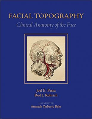
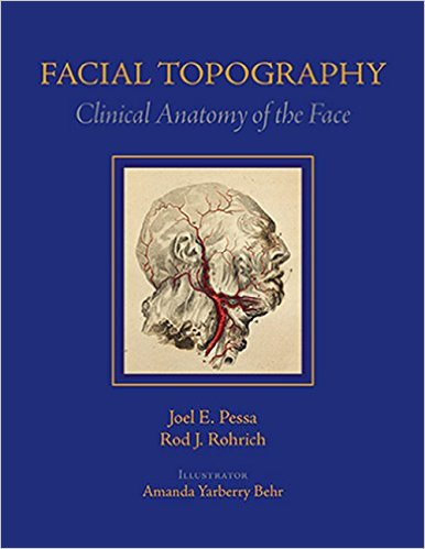
 Authors: Joel E Pessa, MD; and Rod J. Rohrich, MD
Illustrator: Amanda Yarberry Behr, MA, CMI
Publisher: CRC Press (Taylor & Francis Group) – 299 pages
Book Review by: Nano Khilnani
This book is uniquely different from others on examining the anatomy of the face for plastic or reconstructive surgery. Unlike other books, this one focuses on surface landmarks such as shapes, contours, creases and lines that are part of what is called facial topography.
Highly experienced plastic and reconstructive surgeons Joel Pessa and Rod Rohrich point out: “Anatomy is not arbitrary: structures show a remarkable consistency between individuals. It is the difference in the shapes of facial structures and their relationship to one another that determine the unique and distinct appearance of each individual.” These shapes are anatomic regions defined by superficial as well as deep boundaries, they further explain in the Preface.
What are some clinical applications of such topographic analysis of the face of a patient? Here are some:
- Knowledge of the facial fat compartments, including how these units atrophy, helps us understand facial aging
- Such knowledge also enables us to know how diseases of the face develop, and what to do to prevent them. For surgeons, it enables them to better plan their surgical objectives.
- For facial rejuvenation and reconstruction, facial topographic analysis enables surgeons to develop minimally-invasive surgical techniques with smaller incisions. Such procedures would involve greater use of fat, fillers, and toxins for shaping the face.
- Fully understanding the topography of a patient’s face enables the surgeon to know where on the face to do surgical manipulation, volume enhancement, and facial contouring.
These and other considerations in facial aesthetic surgery are presented and discussed in this not-too-large book, highly-focused book of less than 300 pages with just eight chapters:
- Terminology of Facial Topography
- The Central Forehead
- The Cheek
- The Eyelids
- The Nose
- The Temporal Fossa
- The Periauricular Region
- The Lips and Chin
One of the principal valuable features of this book is the use of numerous and fairly good-sized, full-color illustrations: both fine-line drawings as well as actual photos of cadavers and patients.
The authors point out: “our conscious emphasis has been to show rather than tell,”
These images provide a rich learning ground for surgical trainees as well as established surgeons, as learning is dynamic and never ends. The illustrator Amanda Behr has done a great job researching and providing such superb images.
The authors Dr. Joel Pessa and Dr. Rod Rohrich state that they had two main goals in mind when writing this book:
- To demonstrate in a clear and straightforward manner how facial anatomy appears in real life.
- To demonstrate how parts of the face are related to one another to help physicians create a three-dimensional model in their mind’s eye.
A closer look at chapter 7, The Periauricular Region (pages 219-250) shows numerous full-color photos of subjects with a variety of injuries and malformed ears with detailed captions. Much more space is devoted to photos than to the text, adhering to the “show rather than tell” approach of the authors.
In short, this chapter makes the following clinical correlations at the end, with images:
- The lateral neck fold is the topographic landmark for the course of the great auricular nerve. This is the danger zone for potential injury to this sensory nerve.
- By not routinely transecting the posterior auricular tendon, the incidence of injury to the posterior branch of the great auricular nerve is significantly decreased.
- Surgical transitioning is of extreme importance when dissecting from posterior to anterior at the lateral neck fold.
- The anterior auricular crease is a reliable landmark for the superficial temporal artery and the anterior auricular branch.
- The lateral neck fold is an example of a fold associated with a fusion zone. Releasing this fusion zone is the surgical maneuver that softens this fold.
There are very few books that focus on facial topography. This is one of them and it is an excellent one on the subject.
This is a unique book whose premise that “the difference in the shapes of facial structures and their relationship to one another determines the unique and distinct appearance of each individual” makes it stand out from the rest on facial aesthetic anatomy and surgery.
Authors:
Joel E Pessa, MD, FACS is Associate Professor in the Department of Plastic Surgery at the University of Texas Southwestern Medical Center in Dallas, Texas.
Rod J. Rohrich, MD, FACS is Professor and Chairman; Crystal Charity Ball Distinguished Chair in Plastic Surgery; Betty and Woodward Warren Chair in Plastic and Reconstructive Surgery; Distinguished Teaching Professor in the Department of Plastic Surgery at the University of Texas Southwestern Medical Center in Dallas, Texas.
Illustrator:
Amanda Yarberry Behr, MA, CMI


