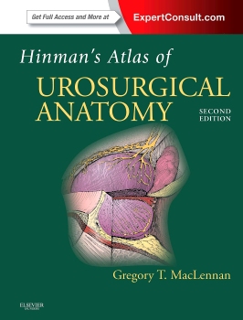
 Author: Gregory T. MacLennan, MD. Illustrated by the late Paul H. Stempen, MA, AMI
Publisher: Elsevier Saunders – 368 pages
Book Review by: Nano Khilnani
This book was first conceived and developed by the late Dr. Frank Hinman Jr. In his Preface the author writes that among Dr. Hinman had these related reasons for coming out with this book:
- To compile information from many sources, including his own studies, for use by urologists
- To create a single, comprehensive, and well-organized textbook that could be consulted quickly and efficiently by urologic surgeons to assist them in planning and performing surgical procedures
This second edition has these improvements over the original edition by Dr. Hinman, namely:
- Colorized anatomical illustrations
- New and relevant images are presented which include clinical and intra-operative photographs from open, laparoscopic and endoscopic procedures, as well as pathologic and radiographic images
- Full-color photos of actual pathology specimens, something that is so rare or almost totally non-existent
Included for you as the purchaser of this print edition of the book is a very user-friendly accompanying website – www.expertconsult.com – which is an online interactive learning platform that presents a vast collection of Elsevier textbook titles with a wide variety of ancillary material.
The website features:
- Fully searchable text
- Integration links that will seamlessly connect you to additional and related content in other ExpertConsult titles
- An image library, with figures that can be easily downloaded into PowerPoint
- Supplementary material such as audio or video clips
If you are already a registered user at ExpertConsult, you can gain access to the online version of this book by going to the above-mentioned website and entering the unique PIN code provided in the scratch-off box on the inside front cover of this book.
If you are a first-time user, do the following:
Register
- Click on Register Now at www.expertconsult.com
- Fill in your user information and click Continue
Activate your book
- Scratch off your Activation Code in the inside front cover of your book
- Enter it into the Enter Activation Code box
- Click Activate Now
- Click the title under My Titles
For technical assistance you can email: [email protected], or call: 800-401-9962 inside the U.S. or 1-314-995-3200 outside the U.S.
The oversized (9×12) format chosen for this book is ideal for presenting visual material of large sizes that provide a lot of important detail. The presentation of ample details is critical in medical texts. The truism of the statement “a picture is worth a thousand words” just cannot be overemphasized in medical education.
Seventeen chapters comprise this book but the organization of the material in them is presented in just three Parts of this book, simply designated as systems, body wall, and organs. The outline below will give you an overview of what you can expect to find in this book:
- Systems: arterial, venous, lymphatic, peripheral nervous, skin, and gastrointestinal tract systems.
- Body Wall: anterolateral body wall; posterolateral and posterior body wall: inguinal region, the pelvis, and the perineum.
- Organs: kidney, ureter, and adrenal glands; bladder, ureterovesical junction, and rectum; prostate and urethral sphincters; female genital tract and urethra; penis and male urethra; and testis.
This wonderful atlas and text created by Dr. Gregory MacLennan provides you detailed illustrations and descriptive text on the anatomy and physiology of organs and systems. This book and its accompanying online resources enable you to see all parts of the genitourinary tract. With it, you will view structures as they appear during surgery. How much better can it get for you?
Gregory T. MacLennan, MD, FRCS (C), FACS, FRCP (C) is Professor of Pathology, Urology and Oncology, and Division Chief in Anatomic Pathology at Case Western Reserve University School of Medicine in the University Hospitals Case Medical Center in Cleveland, Ohio.


