 Authors: Simone Waldt, MD, and Klaus Woertler, MD. With contributions by Matthias Eiber
Publisher: Thieme – 214 pages
Book Review by: Nano Khilnani
Orthopedics is a medical specialty that requires of practitioners to remember a bewildering number and variety of measurement techniques, classification systems, scoring methods, and reference values, the editors write in their Preface.
The techniques of measurement, the systems of classification, the methods of scoring, and the myriad values are so numerous that radiologists, orthopedists, and trauma surgeons just cannot commit all that information and knowledge into their memories
The problem these physicians had faced was that they would have to refer to multiple texts to get all the necessary information required for surgery and treatment. Now, all the fragmented bits and pieces of information on musculoskeletal radiologic measurements and classification systems have been gathered and placed into this single, handy reference guide.
This reference work is ideal for the medical professionals we mention above, as they use radiologic measurements and classifications quite frequently. It can help them make informed diagnoses, and choose the best treatment options.
The book covers a lot of ground, not only in terms of anatomical areas, but also diseases, surgical procedures and operating techniques. It certainly does not provide detailed information that you could find in a full-length textbook, but it functions as a practical handbook. Here below is what this book covers, as we present to you the titles of its chapters:
- Lower Limb Alignment
- Hip
- Knee
- Foot
- Shoulder Joint
- Elbow Joint
- Wrist and Hand
- Spine
- Cranio-cervical Junction and Cervical Spine
- Musculoskeletal Tumors
- Osteoporosis
- Osteoarthritis
- Articular Cartilage
- Hemophilia
- Rheumatoid Arthritis
- Muscle Injuries
- Skeletal Age
The benefits of owning this book are numerous. This book essentially:
- Gathers all currently fragmented information on musculoskeletal radiology measurements and classification system (excluding bone fractures) into one convenient text, eliminating the need for time-consuming memorization and literature searches
- Covers all established measurement methods, using conventional and sectional imaging techniques
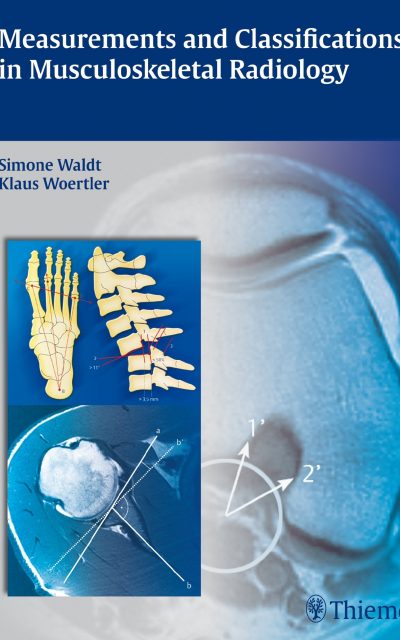
- Includes more than 400 detailed illustrations and radiological images showing reference lines, markings, orientations, and angles, with key values indicated throughout
- Logically structured by anatomic sites and pathologies for easy access to information, with clinical pearls highlighted in every chapter
- Offers practical guidance on the relevance and reliability of each imaging method, preferred techniques for specific diseases, and tips for achieving the clearest and most precise measurement results.
The short, concise chapters are packed with information. They are not wordy and provide you plenty of images, such as charts, drawings, radiographs. They also present text boxes and tables, with data and points that you can learn from at a quick glance. The chapters also have color-coded, numbered tabs at the edges of the pages, so you can quickly turn to the pages and get the information you need in a short time.
This is a well-organized compilation of essential information you need in your busy practice as an orthopedist, radiologist, or surgeon. Drs. Waldt and Woertler have done a marvelous job in providing you this valuable reference source.
Authors:
Simone Waldt, MD is Associate Professor in the Department of Radiology at Klinikum rechts der Isar der Technischen Universitat Munchen in Munich, Germany,
Klaus Woertler, MD is Professor in the Department of Radiology at Klinikum rechts der Isar der Technischen Universitat Munchen in Munich, Germany.


