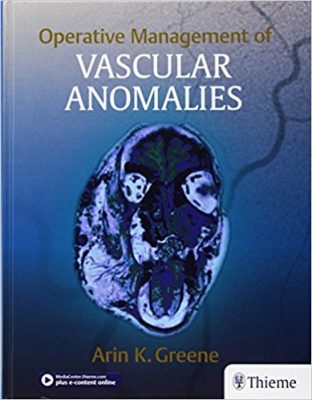
 Editor: Arin K. Greene, MD
Publisher: Thieme – 262 pages
Book Review by: Nano Khilnani
About one in five people are affected by anomalies in their blood vessels and this can cause significant morbidity, writes the editor Dr. Arin Greene. He was prompted to write this guide on vascular abnormalities by being asked by students in his classes, questions such as:
- “What are your surgical indications?”
- “When do you intervene?”
- “How do you perform the procedure?”
- “How do you reduce blood loss?”
- “Where do you place your incision?”
- “What sutures do you use?”
- “How do you manage the patient postoperatively?”
- “What are the complications and how do you treat them?”
He points out that there are no fixed ways of treating what you observe. Something you see on a patient’s lip for instance, may be a manifestation of a lesion that is malignant. If it is, it must be excised, and a lip resection needs to be done.
He writes: “operative techniques for lip conditions usually describe vertical extirpations of malignant lesions, By contrast benign labial vascular anomalies do not require complete resection and can be removed using different methods, such as transverse mucosal excision.”
This work contains many and varied case examples, with a large number of illustrations showing a wide range of malformations and tumors. Tables are used to summarize and differentiate various conditions, including presentation of correct as well as incorrect terms.
For example Table 1.1 on page 4 – Incorrect Terminology Used to Describe Vascular Anomalies – presents the correct biologic names of four types of tumors and four types of malformations. It shows that the tumor infantile hemangioma (the correct biologic name) is sometimes incorrectly referred to as ‘strawberry hemangioma,’ or ‘capillary hemangioma,’ and still other times as ‘cavernous hemangioma’.
It also shows that capillary malformation (correct biologic name) is incorrectly referred to sometimes as ‘port-wine stain’ and sometimes as ‘capillary hemangioma,’ whereas it is actually a tumor. The point is that using an incorrect term can cause confusion, result in the wrong treatment option, or complications.
Table 1.2 – Current Classification of Vascular Anomalies – shown on the following page (5) reflects the most recent (2015) biologic classification of vascular anomalies based on their clinical behavior and cellular characteristics. By using this updated classification, clinicians can diagnose as much as 90 percent of lesions by history and physical examination.
This is an important classification to learn and understand thoroughly because it shows tumors on the left, and malformations on the right. Each type of anomaly (tumor or malformation) is further classified horizontally according to whether it is simple or combined, what types of condition it is (aneurysm, stenosis, etc.), other anomalies it is associated with, and what is unclassified.
Vertically, this table classifies each type of anomaly and tumor based on whether it is benign, locally aggressive or malignant. Also, what types of blood vessels (capillaries, veins, arteries) or location (e/g/lymph) it is found in, and various other characteristics.
Thirteen pediatric cerebrovascular, oral, orthopedic, maxillofacial, plastic, and vascular surgeons and professors in these types of surgeries authored the 16 chapters of this book that has a special focus on malformations and tumors in blood vessels. All of the contributors of content are in Massachusetts, except one each in Maryland and New Jersey.
We list all the titles of Parts and chapters below to give you an overview of the contents of this book:
- Part I. Introduction
- Terminology and Classification
- Principles of Management
- Perioperative Hematologic Management
- Part II. Vascular Tumors
- Infantile Hemangioma
- Pyogenic Granuloma
- Rare Vascular Tumors
- Part III. Vascular Malformations
- Capillary Malformations
- Lymphatic Malformations
- Venous Malformations
- Arteriovenous Malformations
- Anatomic Considerations
- Central Nervous System
- Craniofacial Bones
- Oral Cavity and Airway
- Chest, Abdomen, and Genitalia
- Head and Upper Extremity
- Vertebral Column and Lower Extremity
You can access the videos in the e-book by going to www.ebookstore.thieme.com/register. First, rub off the grey film on the inside front cover of this book, and look at the code revealed therein. Then, click on My E-Books link, select Redeem Access Code, and finally enter that code to claim your e-book. You can then read your e-book online. There are also nine videos for you to see and learn about:
Online Videos:
4-1. Infantile Hemangioma
5-1. Pyogenic Granuloma
6-1. Rare Tumor
7-1. Capillary Malformation
8-1. Lymphatic Malformation
9-1. Venous Malformation
10-1. Arteriovenous Malformation
11-1. Craniofacial Bones
16-1. Intraarticular Venous Malformation
This is a good, comprehensive guide to vascular anomalies with detailed information as to their classification, location, behavior, and other characteristics. It contains 800+ images to make your learning easier and more retentive.
Editor:
Arin K. Greene, MD, MMSc. is affiliated with the Department of Plastic and Oral Surgery at Boston Children’s Hospital, and is Associate Professor of Surgery at Harvard Medical School in Boston, Massachusetts.