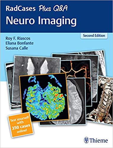
 Editors: Roy F. Riascos MD, Eliana Bonfante MD, and Susana Calle MD
Series Editors: Jonathan M. Lorenz MD and Hector Ferral MD
Publisher: Thieme – 252 pages + 776 illustrations
Book Review by: Nano Khilnani
This book has no table of contents. Instead, it contains 100 carefully selected neuroradiology cases of actual patients for study by radiology residents. This second edition not only adds information found in the first one, it also presents images obtained through additional modalities including 3D reconstructions, nuclear medicine, perfusion, and spectroscopy.
Part of, or besides the 100 cases available in this print edition, you can access 350 cases online! Go to: http://Radcases.Thieme.com. Then once there, type in the access code available on the inside front cover of this print book.
The series editors Drs. Lorenz and Ferral write in their Series Preface that their purpose in publishing this second edition is to assist you to expand your knowledge of, and training in radiology with these new cases, rather than replace the ones that appeared in the first edition.
Meanwhile the editors Drs. Riascos, Bonfante and Calle point out that in the Preface that this book and online resource is intended to maximize your retention of material by providing you questions to answer on the cases presented.
They write: “Our advice is to truly analyze each case as if it were presented to you at your institution of practice. As always, our patients are our best teachers. Try to extract the greatest amount of information from each image and resist the urge to quickly turn the page and read the provided material.”
“The flow of cases intends to mimic what is encountered at the reading station with the added benefit of a third party signaling which images are relevant to the diagnosis A short and concise clinical presentation is provided.”
What is the essence of learning in radiology, have you asked yourself?
The editors state that the essence is the exercise of developing a working diagnosis based on a presented case. This is what “will truly enhance the review process,” they add.
This second edition is different from its predecessor the first edition mainly in that it is interactive. Its main interactivity feature is the questions and answers provided with each of the 100 cases presented in the print version of this book.
Let’s take a look at:
Case 1. Clinical Presentation – A 6 year-old boy presents with headaches (pages 1 and 2).
It begins with three radiographs (A,B,C) shown on page 1. Page 2 begins presentation of those same radiographs with the following captions:
Image A – Sagittal T1 shows peg-shaped cerebellar tonsils lying > 5mm below the level of the foramen magnum, at the level of the posterior arch. Of C1 (black arrow). There is associated dilation of the central canal of the cervical cord (white arrow).
Image B – Axial T2-weighted image demonstrates crowding of the foramen magnum with effacement of the subarachnoid space in the carniocervical junction (white arrow) at
Image C – Transverse gated phase-contrast magnetic resonance images (cerebrospinal fluid study) during the systolic phase and diastolic phase demonstrate lack of flow-related signal and the foramen magnum during systoles (black arrows). During the diastolic phase, there is patent flow ventrally at the foramen magnum (black arrowhead) with obstruction to flow dorsally due to the herniated tonsils (white arrowhead).
Page 2 continues by presenting four sets of headings:
- Differential Diagnosis
- Essential Facts
- Other Imaging Findings
- Pearls and Pitfalls
Each set of headings contains additional points that the resident must look at, to come to the conclusion of what caused the patient to have headaches, and what condition he is in. The Differential Diagnosis presents three possibilities: Chiari I malformation, intracranial hypertension, or tonsillar ectopia.
This is a book that enables neuroradiology residents to look at images, analyze what is shown in them, and come to conclusions on the exact conditions of their patients. It chief value is in assisting them to add to their knowledge of diseases and disorders and at the same time becoming more proficient in their analytical abilities.
Editors:
Roy F. Riascos MD is professor of radiology and chief of neuroradiology in the department of diagnostic and interventional imaging at the University of Texas Health Science Center at Houston in Houston, Texas.
Eliana Bonfante MD is associate professor of radiology in the department of diagnostic and interventional imaging at the University of Texas Health Science Center at Houston in Houston,
Susana Calle MD is staff neuroradiologist in the department of diagnostic and interventional imaging at the University of Texas Health Science Center at Houston in Houston, Texas.
Series Editors:
Jonathan M. Lorenz MD is professor of radiology in the section of interventional radiology at the University of Chicago in Chicago, Illinois.
Hector Ferral MD is senior medical educator at NorthShore University Healthsystem in Evanston, Illinois.