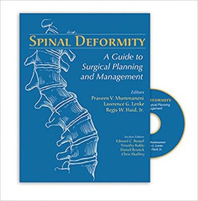
 Editors: Praveen N. Mummaneni, MD; Lawrence G. Lenke, MD; and Regis W. Haid, Jr., MD
Publisher: Quality Medical Publishing – 826 pages
Book Review by: Nano Khilnani
Like many medical and surgical specialties, the field of spinal deformity surgery has been improving, with the advancement of technology, particularly with better imaging techniques and instrumentation. These advances have allowed for greater degrees of spinal deformity correction and lower risk of injury to the spinal cord. Surgeons have also developed more skills, contributing to improved outcomes for patients, the editors point out in the Preface.
Seventy-eight specialists in neurosurgery, orthopedic surgery, spine surgery, and related fields, from all over the United States and three other countries – Canada, Germany, and Turkey – authored the 38 chapters of this large book of over 800 pages published in 2008. All the material in the form of text and graphics is organized into four Parts, namely:
- Part I. Advances in Spine Technology
- Part II. Cervical Spine
- Part III. Thoracic Spine
- Part IV. Lumbosacral Spine
The topics covered are numerous. We name below what is covered in each Part of this book:
Advances in Spine Technology: biomechanics of the spine, conservative management of thoracolumbar deformity, image-guided spinal navigation, interbody fixation options for spinal deformity, minimally-invasive approaches to the cervical spine, spinal biologic, the Lenke classification of adolescent idiopathic scoliosis, and thoracoscopy for anterior thoracic spinal access.
Cervical Spine: basics of cervical kyphosis correction, cervical ankylosing spondylitis, cervical luminoplasty, cervical osteotomy for cervical kyphosis: anterior and posterior options, fixation options for the cervicothoracic junction, fixation options in the occipitocervical junction, and multilevel anterior cervical discectomy and fusion versus corpectomy for cervical kyphosis.
Thoracic Spine: adolescent idiopathic scoliosis: posterior correction techniques, anterior minimally invasive thoracolumbar discectomy and corpectomy techniques, anterior minimally-invasive thoracolumbar junction kyphotic deformity, correction of posttraumatic thoracolumbar junction kyphotic deformity, Scheuermann kyphosis, thoracic Smith-Peterson osteotomy versus pedicle subtraction osteotomy for posterior-only treatment of thoracic kyphosis, thoracoscopy and anterior correction of adolescent idiopathic scoliosis, thoracoscopy for correction of adult thoracic scoliosis, and trajectory techniques of thoracic pedicle screw fixation: anatomc versus straightforward
Lumbosacral Spine: anterior and superior lumbosacral fixation options, high-grade and low-grade spondylolisthesis, indications for extension of lumbar instrumentation to the sacrum, lumbar interbody support options, lumbar pedicle subtraction osteotomy, mini-open posterior lumbar interbody fusion for deformity, sacropelvic fixation options, spondyloptosis, thoracolumbar deformity, and surgical management of lumbar degenerative disease.
The chapters provide materials in a well-organized manner, with a good balance of text, images, tables with data and bullet points, such as step-by-step surgical procedures. Let’s take a look at what you will find in chapter 2, Basics of Cervical Kyphosis Correction, authored by Barunashish Brahma and Frank La Marca.
Kyphosis is exaggerated outward curvature of the spine resulting in a rounded upper back. This chapter provides its material in the following order of coverage of topics:
- Causes of Cervical Kyphosis (Textual discussion)
- Box 12:1 : Causes of Kyphosis
- Iatrogenic : postlaminectomy syndrome, post-irradiation
- Degenerative disc disease
- Congenital deformity
- Tumor
- Infection
- Trauma
- Neuromuscular: neurofibromatosis, denervation of the posterior spinal musculature
- Indications and Contraindications
- Box 12:12: Indications and Contraindications to Surgical Correction of Cervical Kyphosis
- Preoperative Assessment
- Preoperative Planning
- Clinical Problem-Solving: Chart
- Surgical Technique: Key Steps of Technique
- Posterior Approach
- Maintain cervical alignment during patient positioning
- Expose the lateral masses
- Using fluoroscopy, locate the cervical pedicles and articular facets
- Perform anterior grafting to permit elongation of the anterior column
- Leave the lateral mass intact to allow purchase of posterior lateral mass screws for segmental instrumentation
- Posterior Approach
- Anterior Approach (steps provided for this, and three photos presented)
- Box: Postoperative Complications of Cervical Kyphosis Correction
- Postoperative Assessment
- Outcomes
- Case Examples: Case 1 presented, with three radiographs
- Conclusion: Surgical Implications
- References
This is an excellent textbook on the specialty of surgery of deformities of the spine. It is authoritative and comprehensive, with contributions from 78 experts in this field led by the three experienced surgeons.
Editors:
Praveen N. Mummaneni, MD is Associate Professor in the Department of Neurosurgery, and Co-Director of the University of California – San Francisco Spine Center in San Francisco, California.
Lawrence G. Lenke, MD is The Jerome J. Gilden Professor of Orthopedic Surgery, and Professor of Neurological Surgery at Washington State University School of Medicine; and Co-Chair of Spinal, Scoliosis, and Reconstructive Surgery, and Chief of Spinal Surgery at Shriners Hospital for Crippled Children in St. Louis, Missouri.
Regis W. Haid, Jr., MD is with Atlanta Brain and Spine Care in Atlanta, Georgia.
Illustrator:
Jason LeVasseur, MFA


