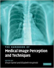
 Editors: Ehsan Samei and Elizabeth Krupinski
Publisher: Cambridge University Press – 424 pages
Book Review by: Nano Khilnani
Medical imaging is a very important feature of medical care these days. The editors write in the initial chapter: “medical images are a core portion of all the information a clinician utilizes to render diagnostic and treatment decisions while a patient is under his or her care.”
Interpretation of medical images is key to diagnosing a patient’s ailment, disorder, or disease. Correct and complete interpretation takes physicians and other health care providers on to the right path of treatment, be it laboratory testing, medication, surgery, or other option.
There are many types of imaging, from the very simple ones to highly detailed ones. Some of the common types and their purposes are:
- Single-projection x-rays such as chest, mammography and musculoskeletal x-rays
- Dynamic x-rays such as three-dimensional computed tomography (CT), fluoroscopy, and magnetic resonance imaging (MRI)
- Nuclear Medicine emission images
- Ultrasound
Recent developments are: digital imaging and multi-detector CT. The range of image types is also growing, with modalities such as molecular imaging and tomosynthesis which are being investigated for various uses such as identifying the margins of lesions during surgical removal to identify cancer cells in the blood..
The variety of medical images is large: They can be
- Acquired with off-the-shelf digital cameras or sophisticated equipment
- Compressed or uncompressed
- Grayscale or color
- Hard copy or soft copy
- Low-resolution to high resolution
The editors point out that while medical imaging is the central technology within radiology, it has also gone beyond radiology and embraced other specialties including cardiology, ophthalmology, pathology, and radiation oncology.
Thirty-eight people mainly in the United States, but also some from the United Kingdom, have contributed content to this book as authors and coauthors of its 31 chapters named below to provide you an overview:
- Medical Image Perception
- Part 1. Historical Reflections and Theoretical Foundations
- A short history of image perception in medical radiology
- Special vision research without noise
- Signal detection theory – a brief history
- Signal detection in radiology
- Lessons from dinners with the giants of modern image science
- Part II. Science of Image Perception
- Perceptual factors in reading medical images
- Cognitive factors in reading medical images
- Satisfaction of search in traditional radiographic imaging
- The role of expertise in radiologic image interpretation
- Image quality and its perceptual relevance
- Beyond the limitations of the human vision system
- Part III. Perception Metrology
- Logistical issues in designing perception experiments
- Receiver operating characteristic analysis: basic concepts and practical applications
- Multireader ROC analysis
- Recent developments in FROC methodology
- Observer models as a surrogate to perception experiments
- Implementation of observer models
- Part IV. Decision Support and Computer Aided Detection
- CAD: An image perception perspective
- Common designs of CAD studies
- Perceptual effect of CAD in reading chest radiographs
- Perceptual issues in mammography and CAD
- How perceptual factors affect the use and accuracy of CAD for interpretation of CT images
- CAD: risks and benefits for radiologists’ decisions
- Part V. Optimization and Practical Issues
- Organization of 2D and 3D radiographic imaging systems
- Applications of AFC methodology in optimization
- Perceptual issues in reading mammograms
- Perceptual optimization of display processing techniques
- Optimization of display systems
- Ergonomic radiologist workspaces in the PACS environment
- Part VI. Epilogue
- Future of medical image perception
A large number of topics are discussed in this book of more than 400 pages. The materials in the chapters are organized systematically so that you can search for information just by reading the chapter title, reading the introductory paragraph(s) to get its gist, looking at the topic headings, look at the tables for data, and view the images for closer understanding. A large number of References are also provided at the end of each chapter for further study and exploration. This is an authoritative, excellent book on medical imaging.
Editors:
Ehsan Samei is a Professor of Radiology, Biomedical Engineering and Physics at Duke University, where he serves as the Director of the Carl E. Ryan Advanced Imaging Laboratories (RAI Labs) and the Director of Graduate Studies for Medical Physics. His current research interests include medical image formation, analysis, display, and perception, with particular focus on quantitative and molecular imaging.
Elizabeth Krupinski is a Professor at the University of Arizona in the Departments of Radiology, Psychology, and Public Health. She is the Associate Director of Evaluation and Assessment for the Arizona Telemedicine Program, President of the Medical Image Perception Society, and serves on the editorial boards of a number of journals in both radiology and telemedicine.


