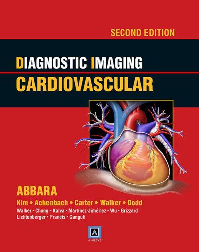
Editors: Suhny Abbarra, MD; Stephan Achenbach, MD; Jonathan R, Dodd, MD; Brett W. Carter, MD; Raymond J. Kim, MD; Chirstopher M. Walker, MD; T. Gregory Walker, MD; Jonathan Hero Chung, MD; Carol C. Wu, MD; John D. Grizzard, MD; John P. Lichtenberger III MD; Sanjeeva P. Kalva, MD; Sanjeev A. Francis, MD; Santiago Martinez-Jimenez, MD; Suvranu “Shoey” Ganguli, MD
Publisher: Wolters Kluwer | Lippincott, Williams & Wilkins (LWW)
Book Review by: Nano Khilnani
This is a massive textbook with 16 sections, 203 chapters (of which 32 are new as compared to the first edition), and nearly 2,500 high-quality images.
And it is an outstanding book because in my opinion, it represents a large and valuable collection of material from top experts in cardiology and radiology; a skillful organization and presentation of contents; excellent quality images; and concise, lucid explanations in its text of conditions diseases, and disorders.
This book represents the work of 15 editors and 21 authors of material who are experts in their respective fields.
Imaging as a diagnostic tool has grown in use extensively in medicine in general, but more so in cardiology in particular. Two relatively newer types of imaging – computed tomography (CT) and cardiac magnetic resonance (CMR) are now widely available, and have expanded use, flexibility, and popularity, writes Dr. Gilbert L. Raff, director of Advanced Cardiovascular Imaging in Rochester, Michigan, in his Foreword to this book.
The longstanding, or what are called “traditional” imaging methods – digital fluoroscopy, echocardiography, and nuclear cardiology – are now evolving into new technologies, with current capabilities having developed further before. The capabilities include: preoperative planning of trans-catheter valve replacement and trans-cutaneous treatment of many congenital diseases.
The scope of this book on imaging of the heart and related blood vessels is, to say the least, vast. Its approach is not just anatomical but also physiological, showing detailed views of the different parts of the human heart as well as different types of diseases, disorders and conditions.
To give you a broad overview of the contents of this book, these are its sections:
- Introduction and Overview
- Congenital
- Shunts
- Valvular
- Pericardial
- Neoplastic
- Cardiomyopathy
- Coronary Artery Disease
- Heart Failure
- Electrophysiology
- Pulmonary Vasculature
- Arterial: a. Thoracic Aorta, and b. Abdominal Aorta and Visceral Vasculature
- Venous
- Extracranial Cerebral Arteries
- Renal Vasculature
- Peripheral Vasculature: a. Introduction and Overview, b. Vasculature of the trunk, and c. Lower Extremity Vasculature
The different types visuals used in this unique book are not only CTs, MRIs and other types of scans, but also full-color drawings labeled with very helpful and informative detailed anatomical descriptions and explanations.
The information in textual form will help you the medical student, resident or practitioner in cardiology or cardiac surgery, with analysis of the problems and selection of possible solutions and treatment options.
For example, in subsection 8 on arterial stenosis in Section 8 – Coronary Artery Disease – you will find broad information on this condition with narrowing of the arterial walls which limits the amount of blood that can flow to and from the heart in a box entitled Key Facts with headings such as Terminology, Imaging, Top Differential Diagnoses, and Clinical Issues.
Viewing the various images of your patient’s stenosis and the extent of his or her occlusion in the damaged artery or arteries can help you assess the severity of his or her condition and make informed, correct medical and surgical decisions in a timely manner. This is absolutely critical.
You can benefit from using the online resources of this book in the following ways:
- Searchable content
- Expanded diagnostic tips and references
- Additional annotated images
To access your Amirsys eBook Advantage features, do the following
- Scratch off the silver coating on inside front cover to reveal the license key
- Go to http://ebooks.amirsys.com
- Existing users: log in to your account
- New users: register for a new account
- Users outside the U.S. need to select “Other” for State
- Register your title with this license key
(You may need to log out and log back in to see your new eBook in your list of registered titles).
Editors:
Suhny Abbarra, MD, FSCCT is Professor of Radiology, Chief of the Division of Cardiac and Thoracic Imaging, and Director of 3D Image Processing Laboratory at University of Texas Southwestern Medical Center in Dallas, Texas.
Stephan Achenbach, MD is Chairman of the Department of Cardiology at University of Erlangen in Erlanger, Germany.
Brett W. Carter, MD is Assistant Professor of Radiology at the University of Texas MD Anderson Cancer Center in Houston, Texas
Jonathan Hero Chung, MD is Associate Professor in the Department of Radiology, Director of Radiology Professional Quality Assurance, and Director of Cardiopulmonary Imaging Fellowship at National Jewish Health Center in Denver, Colorado.
Jonathan R, Dodd, MD, MSc, MRCPI, FFR (RCSI) is Associate Professor of Radiology at University College Dublin, and Director of Radiology at St. Vincent’s University Hospital in Dublin, Ireland.
Sanjeev A. Francis, MD is Director of the Cardio-Oncology Program and the Cardio-MRI/CT Program, and Instructor in Medicine at Harvard Medical School and Massachusetts General Hospital in Boston, Massachusetts. Suvranu “Shoey” Ganguli, MD is Assistant Professor of Radiology at Harvard Medical School and Vascular & Interventional Radiology at Massachusetts General Hospital in Boston, Massachusetts.
John D. Grizzard, MD is Associate Professor of Radiology and Section Chief of Non-Invasive Cardiovascular Imaging at VCU Health Systems in Richmond, Virginia.
Sanjeeva P. Kalva, MBBS, MD, FSIR is Associate Professor of Radiology and Chief of the Division of Interventional Radiology at University of Texas Southwestern Medical Center in Dallas, Texas.
Raymond J. Kim, MD is Director of Duke Cardiovascular Magnetic Resonance Center and Professor of Medicine and Radiology at Duke University Medical School in Durham, North Carolina.
John P. Lichtenberger III, MD is Chief of Cardiothoracic Imaging at David Grant Medical Center at Travis Air Force Base in California. He is also Assistant Professor of Radiology at Uniformed Services University of Health Sciences in Bethesda, Maryland.
Santiago Martinez-Jimenez, MD is Associate Professor of Radiology at the University of Missouri – Kansas City, and Saint Luke’s Hospital of Kansas City in Kansas City, Missouri.
Christopher M. Walker, MD is Assistant Professor of Radiology at Saint Luke’s Hospital of Kansas City and the University of Missouri in Kansas City, Missouri.
T. Gregory Walker, MD, FSIR is Assistant Professor of Radiology at Harvard Medical School, and Associate Director of Fellowship in the Division of Interventional Radiology at Massachusetts General Hospital in Boston, Massachusetts.
Carol C. Wu, MD is Instructor of Radiology at Harvard Medical School and Assistant Radiologist at Massachusetts General Hospital in Boston, Massachusetts.
Contributing Authors:
Gerald F. Abbott, MD
Lowie M.R. Van Assche, MD
Darragh Brady, MD, MRCPI
Roy Bryan, MD, MBA
Rebecca S. Cornelius, MD, FACR
Daniel W. Entrikin, MD
Gudrun Feuchtner, MD
Andrew J. Gunn, MD
Bronwyn E. Hamilton, MD
Cameron Hassani, MD
Terrance T. Healey, MD
Michael T. Lu, MD
Jeffrey P. Kanne, MD
Ali Devrim Karaosmanoglu, MD
Naveen M. Kulkarni, MD
Kathryn M. Olsen, MD
C. Douglas Phillips, MD, FACR
Carlos A. Rojas, MD
Melissa L. Rosado-de-Christensen, MD, FACR
Rahul Sheth, MD
Tyler H. Ternes, MD


