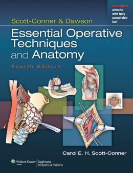
 Editor: Carol. E.H. Scott-Conner, MD, PhD, MBA
Publisher: Wolters Kluwer | Lippincott, Williams & Wilkins – 751 pages
Book Review by: Nano Khilnani
The need of general surgeons for precise anatomical knowledge of the entire human body has increased over the years, and more so for specialist surgeons who operate on certain regions. These were the reasons for the publication of original version of this book, as mentioned by Dr. James Hardy who wrote the Foreword to the first edition
Those reasons remain for this the fifth edition, but a lot of other needs have arisen over the years as this book has evolved and improved. Its editor Carol E.H. Scott-Conner writes about what is new in this current edition, and among other items, they are:
- This entire book has been structured around the 2013-14 curriculum outline of the Surgical Council on Resident Education (SCORE).
- While the “essential common” and “essential uncommon” procedures remain in the print edition, the “complex” procedures have been moved to the web edition.
- The first edition had 72 chapters; the current one, 134, an 86 percent increase.
- The number of procedures, color photos, and even authors has grown.
Notwithstanding the changes – some of which we mention above – good features have been retained. The chapters still start with a cream-colored page that lists Steps in Procedure, List of Structures, and Hallmark Anatomic Considerations. These specific lists for each type of surgical procedure in the various chapters are meant for a quick review before the operation begins.
For example in chapter 128- Below-Knee Amputation – of Section VI – Lower Extremity:
The first paragraph in this chapter advises the surgeon: “Most amputations are performed for ischemia (deficient supply of blood to the body part). The choice of the level of amputation requires mature judgment. Although it is important to salvage as much length as possible, a poor initial choice of level may doom the patient to a second amputation, often at a significantly higher level.”
The Steps in Procedure are numerous, but these are a few of them: Skin incision should provide longer posterior flap than anterior flap…Preserve as much length as possible…Divide all soft tissues to tibia anteriorly and through fascia of muscles laterally…Divide tibia 1 to 2 cm above level of skin incision…Smooth ends of bone…
The Hallmark Anatomic Complications are two: Ischemia of stump, and Neuroma formation. Neuroma is a mass of nerve tissue in an amputation stump resulting from abnormal re-growth of the stumps of severed nerves.
The List of Structures mentions names of various arteries, bones, fascia, muscles, nerves, veins, and other parts that play roles in the functioning of the leg.
52 contributors from the United States (of which more than two-thirds or 35 are from Iowa) plus Iceland, and Lebanon wrote the 134 chapters of this extensive book on anatomy and operative techniques.
This book comes with access to the complete contents online, full searchable, plus other valuable features. Scratch off the gray sticker on the inside front cover of your book to get your access code. Three simple steps to gain instant access.
1) Visit http://solution.lww.com
2) Enter your access code
3) Follow the instructions to activate your access
For technical assistance, call 1-800-468-1128 in the United States or 1-410-528-4000 outside the U.S., or email: [email protected]
The large number of chapters – 134 – and the large range of techniques covered in this book can be overwhelming, so the material is organized around six broad sections:
- The Head and Neck
- The Pectoral Region and the Chest
- The Upper Extremity
- The Abdominal Region
- The Sacral Region and the Perineum
- The Lower Extremity
Numerous multicolor images (actual photographs of procedures as well as sketches in color) make learning much easier than simply reading the text or even looking at black-and-white drawings.
Two interesting features in this book are Anatomical Points and Technical Points.
In chapter 128 as we referenced above, the Anatomical Points mentions, among other guidelines, “The division of the tibia about four fingerbreadths inferior to the tibial tuberosity corresponds to approximately the level of the greatest circumference of the leg. At this location, the greater saphenous vein and accompanying saphenous nerve are located in the superficial fascia just posterior to the medial border of the tibia – that is, in the fascia overlaying the tibial origin of the soleus muscle.”
In the same chapter the Technical Points begins to inform the reader: “Plan a skin incision with a long posterior flap. The length of the posterior flap should approximate the: transverse diameter of the leg. As extra length of flap can always be trimmed, it is advisable to make the flap too long at the initial incision.”
Carol E.H. Scott-Conner, MD, PhD, MBA is Professor of Surgery in the Department of Surgery at the University of Iowa Roy J. and Lucille A. Carver School of Medicine in Iowa City, Iowa.