 Authors: Edward C. Weber, DO, Joel A. Vilensky, PhD, Stephen W. Carmichael, PhD, DSc, and Kenneth C. Lee, MD
Publisher: Elsevier Saunders – 519 pages
Book Review by: Nano Khilnani
You can gain more value by accessing additional information and knowledge relating to the topics covered in this book by going to: www.studentconsult.com. Among other features available to you, you can do the following:
- Access the full text online
- Perform rapid searches on any topic
- Add your own notes and bookmarks.
Go to the inside front cover of this book to scratch off a box here that provides you with a PIN code. Here are the instructions relating to the use of your PIN code:
Login or Sign Up at www.StudentConsult.com
- Scratch off your PIN code from the box on the inside front cover of this book
- Enter PIN code into the Redeem a Book Code box on this website
- Click Redeem
- Go to My Library
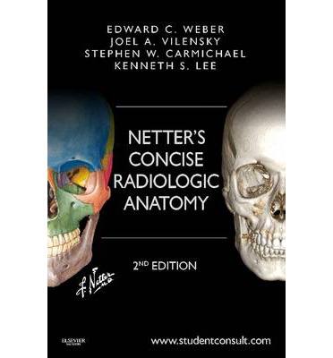
This book compares radiographic images (computer tomography or CT and magnetic resonance imaging or MRI) with the excellent, detailed images of the late renowned medical illustrator Frank H. Netter, MD, making learning anatomy of various parts of the human body easier and more useful for you the student.
This comparison approach is demonstrated in hundreds of instances in this book with Dr. Netter’s full-color drawings on the left and the radiographs on the right of each page. It is quite comprehensive in coverage with several sections in this book that cover
- Head and Neck
- Back and Spinal Cord
- Thorax
- Abdomen
- Pelvis and Perineum
- Upper Limb
- Lower Limb
This book contains about a thousand images showing detailed representations of the myriad portions of the seven sections of our body listed above. The Netter illustrations are in full color, while the radiographic images are mainly black and white. Some of the radiographs however are in color and surprisingly clear and detailed.
At the bottom of each full-color Netter drawing is a Clinical Note that is helpful to you in terms of what you need to know and do.
For example, the Clinical Note in the depiction of Brainstem and Vertebral Arteries on page 118 in the Head and Neck section states: “The absence of a flow void (indicating blood flow) on the MR image may be direct evidence of arterial occlusion. Significant discrepancy in the size of normal vertebral arteries is common and no clinical significance. Vertebrobasilar artery insufficiency often presents with neurologic dysfunction that is clinically distinct from the more common carotid artery disease.”
The team of two specialists each in radiology and anatomy named below has done an outstanding job of putting together this handy guide to anatomic images of various parts of the human anatomy. This is a unique, valuable addition to your medical library.
Edward C. Weber, DO is Radiologist at The Imaging Center in Fort Wayne, Indiana; Consultant at Medical Clinic of Big Sky in Big Sky, Montana; Adjunct Professor of Anatomy and Cell Biology, and Volunteer Clinical Professor of Radiology and Imaging Sciences at Indiana University School of Medicine in Fort Wayne, Indiana.
Joel A. Vilensky, PhD is Professor of Anatomy and Cell Biology at Indiana University School of Medicine in Fort Wayne, Indiana.
Stephen W. Carmichael, PhD, DSc is Editor Emeritus of Clinical Anatomy; Professor Emeritus of Anatomy, and Professor Emeritus of Orthopedic Surgery at Mayo Clinic in Rochester, Minnesota.
Kenneth S. Lee, MD is Associate Professor of Radiology, Director of Musculoskeletal Ultrasound, and Medical Director of Transitional Imaging, at University of Wisconsin School of Medicine and Public Health in Madison, Wisconsin.
Frank H. Netter, MD was acknowledged as one of the finest and most medical illustrators in the world. His 13-book Netter Collection of Medical Illustrations, which includes the greater part of more than 20,000 paintings, became and remains one of most famous medical works every published. Dr. Netter was born in 1906 in New York City. He studied art at the Art Students League and the National Academy of Design before entering medical school at New York University, where he received his medical degree in 1931. After establishing a surgical practice in 1933, he continued illustrating as a sideline, which he had begun doing as a medical student. He ultimately gave up his practice in favor of a full-time commitment to art. He died in 1991 leaving a rich legacy of intellectually valuable medical illustrations.


