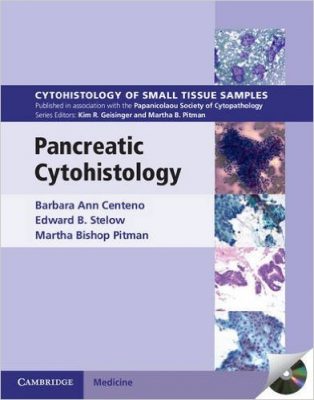
 (Part of the Series: Cytohistology of Small Tissue Samples)
Editors: Barbara Ann Centeno, MD; Edward B. Stelow, MD; and Martha Bishop Pitman, MD
Publisher: Cambridge University Press – 175 pages
Book Review by: Nano Khilnani
There are several diseases of the pancreas, according to a Wikipedia article. Some of them are:
- Pancreatitis (acute and chronic types)
- Diabetes mellitus: type 1, with insufficient insulin synthesis and secretion, and type 2, insulin resistance, resulting in failure of pancreas to match insulin production to demand)
- Exocrene pancreatic insufficiency
- Cystic fibrosis: tissue scarring and cyst formation
- Pancreatic pseudocysts: collections of fluids rich in amylase, enzymes, necrotic tissue
- Cysts: common in the pancreas, and often benign
- Congenital malformations (pancreas divisum and annular pancreas)
- Neoplasms (benign and predisposed to tumors, e.g. Zollinger-Ellison syndrome)
- Hemosuccus pancreaticus
This book discusses these diseases and the process of examining tissue samples to discover disease, disorders, abnormalities, and anomalies. The editors named above and four other physicians named at the end of this brief book review have written this relatively brief book yet packed with a lot of information in text and graphic form that succinctly covers and discusses several areas within pancreatic cytohistology.
An overview of the coverage of this text is provided below with the list of its eleven chapters:
- Introduction: Principles and practices for small tissue samples in the diagnosis of pancreatic masses
- Endoscopic ultrasound-guided fine-needle aspiration of the pancreas
- Tissue handling and processing
- Ancillary studies
- Cytology and histology of the normal pancreas, and contaminants
- Non-neoplastic mass lesions
- Cystic lesions
- Pancreatic and ductal adenocarcinoma and variants
- Solid nonductal epithelial neoplasia
- Secondary neoplasia (metastases, direct extension of the adjacent tumors, and the lymphoreticular lesions
- Mesenchymal neoplasia
The editors provide good coverage of the subjects listed above, and an examination of a sample chapter is provided here. Let us take a look at the outline and images provided in chapter 8 entitled Pancreatic Ductal Adenocarcinoma and Variants.
- Introduction
- Clinical Features
- Radiographic Features
- Pancreatic Cancer Precursors
- Cytological features
- Histological features
- Ancillary testing and differential diagnosis
- Ductal Adenocarcinoma
- Undifferentiated (Anaplastic) Adenocarcinoma
- Undifferentiated Carcinoma with Osteoclast-Like Giant Cells
- Adenosquamous Carcinoma
- Colloid (Mucinous Noncystic) Carcinoma
- Signet Ring Cell Carcinoma
- Oncocytic Carcinoma
- Medullary Carcinoma
- Clinical Vignettes
- Case 1: 73-year-old man being followed for urethral carcinoma
- Case 2: 71-year-old woman with abdominal pain and mass in the pancreas
- References : separate lists for Introduction and other types of carcinomas
For each of the types of carcinoma listed above in bold letters above, the authors of this chapter discuss the cytological and histological features, ancillary testing and differential diagnosis.
This is an excellent work on pancreatic cytohistology. It is concise, focusing on the proper methods of tissue examination, analysis, and interpretation. It is very useful with numerous micrographs enabling the reader to imbue a lot of detailed information about diseases and disorders in the pancreas. It is a must for the pancreas specialist.
Editors:
Barbara Ann Centeno, MD is Vice Chair of Clinical Services, Assistant Chief, Pathology Service, Director of Cytopathology, and Senior Member of the Department of Anatomic Pathology at H.Lee Moffitt Cancer Center and Research Institute; and Professor of Clinical Oncologic Sciences at Morsani College of Medicine at the University of South Florida in Tampa, Florida.
Edward B. Stelow, MD is Associate Professor in the Department of Pathology at the University of Virginia in Charlottesville, Virginia.
Martha Bishop Pitman, MD is Medical Director of the Cytopathology Laboratory at Massachusetts General Hospital; and Associate Professor of Pathology at Harvard Medical School in Boston, Massachusetts.
Contributing Authors:
William R. Brugge, MD
Junsung Choi, MD
Abdurrahman Kadayifci, MD
Brian Morse, MD